 Explore
 Validate
 Learn
Learn Western blot
 Immunocytochemistry
Antibody data
- Antibody Data
- Antigen structure
- References [3]
- Comments [0]
- Validations
- Western blot [1]
- Immunohistochemistry [1]
Submit
Validation data
Reference
Comment
Report error
- Product number
- NB100-1920 - Provider product page

- Provider
- Novus Biologicals
- Proper citation
- Novus Cat#NB100-1920, RRID:AB_10001381
- Product name
- Rabbit Polyclonal Nucleolin Antibody
- Antibody type
- Polyclonal
- Description
- Unpurified.
- Reactivity
- Human, Rat
- Host
- Rabbit
- Isotype
- IgG
- Vial size
- 0.1 ml
- Storage
- Aliquot and store at -20C or -80C. Avoid freeze-thaw cycles.
Submitted references The Zinc-Finger Antiviral Protein ZAP Inhibits LINE and Alu Retrotransposition.
Recognition and binding of the human selenocysteine insertion sequence by nucleolin.
Recognition and binding of the human selenocysteine insertion sequence by nucleolin.
Moldovan JB, Moran JV
PLoS genetics 2015 May;11(5):e1005121
PLoS genetics 2015 May;11(5):e1005121
Recognition and binding of the human selenocysteine insertion sequence by nucleolin.
Wu R, Shen Q, Newburger PE
Journal of cellular biochemistry 2000 Apr;77(3):507-16
Journal of cellular biochemistry 2000 Apr;77(3):507-16
Recognition and binding of the human selenocysteine insertion sequence by nucleolin.
Wu R, Shen Q, Newburger PE
Journal of cellular biochemistry 2000 Apr;77(3):507-16
Journal of cellular biochemistry 2000 Apr;77(3):507-16
No comments: Submit comment
Supportive validation
- Submitted by
- Novus Biologicals (provider)
- Main image

- Experimental details
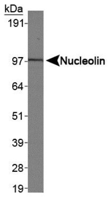
- Western Blot: Nucleolin Antibody [NB100-1920] - Analysis of Nucleolin in HeLa nuclear extracts.
Supportive validation
- Submitted by
- Novus Biologicals (provider)
- Main image
- Experimental details
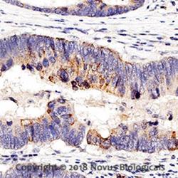
- Immunohistochemistry-Paraffin: Nucleolin Antibody [NB100-1920] - IHC analysis of a formalin fixed paraffin-embedded (FFPE) human colon cancer using 1:750 conc. of Nucleolin antibody on a Bond Rx autostainer (Leica Biosystems). The assay involved 20 minutes of heat induced antigen retrieval (HIER) using 10mM sodium citrate buffer (pH 6.0) and endogenous peroxidase quenching with peroxide block. The sections were incubated with primary antibody for 30 minutes and Bond Polymer Refine Detection (Leica Biosystems) with DAB was used for signal development followed by counterstaining with hematoxylin. Whole slide scanning and capturing of representative images (20X) was performed using Aperio AT2 (Leica Biosystems). Cytoplasmic staining was observed in normal cells and nuclear staining was observed in cancer cells. Staining was performed by Histowiz.
