Explore
 Validate
 Learn
Learn Western blot
Antibody data
- Antibody Data
- Antigen structure
- References [14]
- Comments [0]
- Validations
- Western blot [5]
- Immunocytochemistry [2]
- Immunohistochemistry [1]
- Flow cytometry [1]
- Other assay [6]
Submit
Validation data
Reference
Comment
Report error
- Product number
- 36-8800 - Provider product page

- Provider
- Invitrogen Antibodies
- Product name
- Phospho-ERK1/ERK2 (Thr202, Tyr204) Polyclonal Antibody
- Antibody type
- Polyclonal
- Antigen
- Synthetic peptide
- Description
- This antibody is predicted to react with Xenopus and zebrafish based on 100% sequence homology.
Submitted references Empagliflozin mitigates type 2 diabetes-associated peripheral neuropathy: a glucose-independent effect through AMPK signaling.
Preventative Effect of Mebendazole against Malignancies in Neurofibromatosis 1.
Mitogen-activated protein kinases MPK3 and MPK6 are required for stem cell maintenance in the Arabidopsis shoot apical meristem.
A Novel Heterozygous Deletion Variant in KLOTHO Gene Leading to Haploinsufficiency and Impairment of Fibroblast Growth Factor 23 Signaling Pathway.
Fc-mediated activity of EGFR x c-Met bispecific antibody JNJ-61186372 enhanced killing of lung cancer cells.
Oncogenic Ras differentially regulates metabolism and anoikis in extracellular matrix-detached cells.
The role of multicellular aggregation in the survival of ErbB2-positive breast cancer cells during extracellular matrix detachment.
Antihypertensive drug Valsartan promotes dendritic spine density by altering AMPA receptor trafficking.
A tetra(ethylene glycol) derivative of benzothiazole aniline enhances Ras-mediated spinogenesis.
The important roles of RET, VEGFR2 and the RAF/MEK/ERK pathway in cancer treatment with sorafenib.
Post-receptor IGF1 insensitivity restricted to the MAPK pathway in a Silver-Russell syndrome patient with hypomethylation at the imprinting control region on chromosome 11.
Growth-factor receptor-bound protein-2 (Grb2) signaling in B cells controls lymphoid follicle organization and germinal center reaction.
Osteopontin is linked to p65 and MMP-9 expression in pulmonary adenocarcinoma but not in malignant pleural mesothelioma.
15-Deoxy-Delta12,14-prostaglandin J2 regulates endogenous Cot MAPK kinase kinase 1 activity induced by lipopolysaccharide.
Abdelkader NF, Elbaset MA, Moustafa PE, Ibrahim SM
Archives of pharmacal research 2022 Jul;45(7):475-493
Archives of pharmacal research 2022 Jul;45(7):475-493
Preventative Effect of Mebendazole against Malignancies in Neurofibromatosis 1.
Staedtke V, Gray-Bethke T, Riggins GJ, Bai RY
Genes 2020 Jul 8;11(7)
Genes 2020 Jul 8;11(7)
Mitogen-activated protein kinases MPK3 and MPK6 are required for stem cell maintenance in the Arabidopsis shoot apical meristem.
Lee H, Jun YS, Cha OK, Sheen J
Plant cell reports 2019 Mar;38(3):311-319
Plant cell reports 2019 Mar;38(3):311-319
A Novel Heterozygous Deletion Variant in KLOTHO Gene Leading to Haploinsufficiency and Impairment of Fibroblast Growth Factor 23 Signaling Pathway.
Martín-Núñez E, Donate-Correa J, Kannengiesser C, De Brauwere DP, Leroy C, Oudin C, Friedlander G, Prieto-Morín C, Tagua VG, Ureña-Torres PA, Navarro-González JF
Journal of clinical medicine 2019 Apr 12;8(4)
Journal of clinical medicine 2019 Apr 12;8(4)
Fc-mediated activity of EGFR x c-Met bispecific antibody JNJ-61186372 enhanced killing of lung cancer cells.
Grugan KD, Dorn K, Jarantow SW, Bushey BS, Pardinas JR, Laquerre S, Moores SL, Chiu ML
mAbs 2017 Jan;9(1):114-126
mAbs 2017 Jan;9(1):114-126
Oncogenic Ras differentially regulates metabolism and anoikis in extracellular matrix-detached cells.
Mason JA, Davison-Versagli CA, Leliaert AK, Pape DJ, McCallister C, Zuo J, Durbin SM, Buchheit CL, Zhang S, Schafer ZT
Cell death and differentiation 2016 Aug;23(8):1271-82
Cell death and differentiation 2016 Aug;23(8):1271-82
The role of multicellular aggregation in the survival of ErbB2-positive breast cancer cells during extracellular matrix detachment.
Rayavarapu RR, Heiden B, Pagani N, Shaw MM, Shuff S, Zhang S, Schafer ZT
The Journal of biological chemistry 2015 Apr 3;290(14):8722-33
The Journal of biological chemistry 2015 Apr 3;290(14):8722-33
Antihypertensive drug Valsartan promotes dendritic spine density by altering AMPA receptor trafficking.
Sohn YI, Lee NJ, Chung A, Saavedra JM, Scott Turner R, Pak DT, Hoe HS
Biochemical and biophysical research communications 2013 Oct 4;439(4):464-70
Biochemical and biophysical research communications 2013 Oct 4;439(4):464-70
A tetra(ethylene glycol) derivative of benzothiazole aniline enhances Ras-mediated spinogenesis.
Megill A, Lee T, DiBattista AM, Song JM, Spitzer MH, Rubinshtein M, Habib LK, Capule CC, Mayer M, Turner RS, Kirkwood A, Yang J, Pak DT, Lee HK, Hoe HS
The Journal of neuroscience : the official journal of the Society for Neuroscience 2013 May 29;33(22):9306-18
The Journal of neuroscience : the official journal of the Society for Neuroscience 2013 May 29;33(22):9306-18
The important roles of RET, VEGFR2 and the RAF/MEK/ERK pathway in cancer treatment with sorafenib.
Mao WF, Shao MH, Gao PT, Ma J, Li HJ, Li GL, Han BH, Yuan CG
Acta pharmacologica Sinica 2012 Oct;33(10):1311-8
Acta pharmacologica Sinica 2012 Oct;33(10):1311-8
Post-receptor IGF1 insensitivity restricted to the MAPK pathway in a Silver-Russell syndrome patient with hypomethylation at the imprinting control region on chromosome 11.
Montenegro LR, Leal AC, Coutinho DC, Valassi HP, Nishi MY, Arnhold IJ, Mendonca BB, Jorge AA
European journal of endocrinology 2012 Mar;166(3):543-50
European journal of endocrinology 2012 Mar;166(3):543-50
Growth-factor receptor-bound protein-2 (Grb2) signaling in B cells controls lymphoid follicle organization and germinal center reaction.
Jang IK, Cronshaw DG, Xie LK, Fang G, Zhang J, Oh H, Fu YX, Gu H, Zou Y
Proceedings of the National Academy of Sciences of the United States of America 2011 May 10;108(19):7926-31
Proceedings of the National Academy of Sciences of the United States of America 2011 May 10;108(19):7926-31
Osteopontin is linked to p65 and MMP-9 expression in pulmonary adenocarcinoma but not in malignant pleural mesothelioma.
Frey AB, Wali A, Pass H, Lonardo F
Histopathology 2007 May;50(6):720-6
Histopathology 2007 May;50(6):720-6
15-Deoxy-Delta12,14-prostaglandin J2 regulates endogenous Cot MAPK kinase kinase 1 activity induced by lipopolysaccharide.
Caivano M, Rodriguez C, Cohen P, Alemany S
The Journal of biological chemistry 2003 Dec 26;278(52):52124-30
The Journal of biological chemistry 2003 Dec 26;278(52):52124-30
No comments: Submit comment
Supportive validation
- Submitted by
- Invitrogen Antibodies (provider)
- Main image

- Experimental details
- Western blot analysis of Phospho-MAPK1/3 pThr202/Tyr204 was performed by loading rat brain lysates onto a SDS-PAGE gel. Proteins were transferred to a membrane, blocked, and probed with a Phospho-MAPK1/3 pThr202/Tyr204 polyclonal antibody (Product # 36-8800) at a dilution of 1:1000, followed by an HRP-conjugated anti-rabbit IgG secondary antibody. Detection was performed using a chemiluminescent substrate.

- Submitted by
- Invitrogen Antibodies (provider)
- Main image
- Experimental details
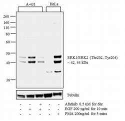
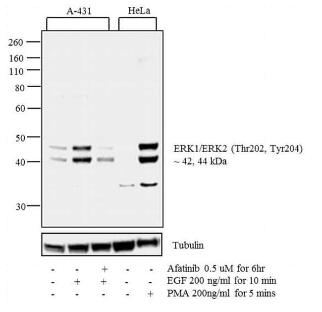
- Western blot analysis was performed on whole cell extracts (30 µg lysate) of A-431 (1), A-431 treated with EGF (200 ng/mL for 10 minutes) (2), A-431 treated with Afatinib followed by EGF (0.5 uM for 6 hours, 200 ng/mL for 10 minutes) (3), HeLa (4) and HeLa treated with PMA (200 ng/mL for 5 min). The blot was probed with Anti-Phospho-ERK1/ERK2 (Thr202, Tyr204) Rabbit Polyclonal Antibody (Product # 36-8800, 1:250 dilution) and detected by chemiluminescence using Goat anti-Rabbit IgG (H+L) Superclonal™ Secondary Antibody, HRP conjugate (Product # A27036, 0.25 µg/mL, 1:4000 dilution). 42, 44 kDa band corresponding to Phospho-ERK1/ERK2 (Thr202, Tyr204) was detected to increase upon EGF and PMA treatments in the cell lines tested. Pre-treatment with EGFR-antagonist, Afatinib, resulted in inhibition of Phospho-ERK1/ERK2 (Thr202, Tyr204) in A-431 cell line. Known quantity of protein samples were electrophoresed using Novex® NuPAGE® 4-12 % Bis-Tris gel (Product # NP0321BOX), XCell SureLock System (Product # EI0002) and Novex Protein Standard (Product # LC5800). Proteins were then transferred onto a nitrocellulose membrane with iBlot® 2 Dry Blotting System (Product # IB21001). The membrane was probed with the relevant primary and secondary Antibody following blocking with 5 % skimmed milk. Chemiluminescent detection was performed using Pierce™ ECL Western Blotting Substrate (Product # 32106).
- Submitted by
- Invitrogen Antibodies (provider)
- Main image

- Experimental details
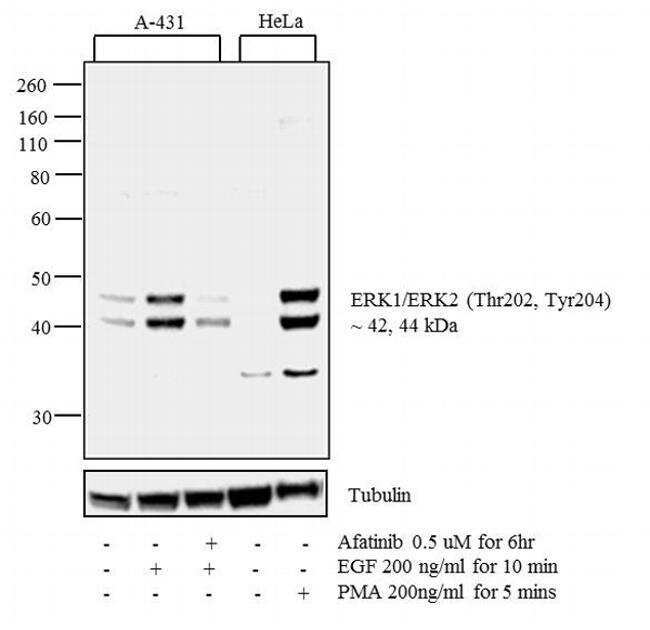
- Western blot analysis was performed on whole cell extracts (30 µg lysate) of A-431 (1), A-431 treated with EGF (200 ng/mL for 10 minutes) (2), A-431 treated with Afatinib followed by EGF (0.5 uM for 6 hours, 200 ng/mL for 10 minutes) (3), HeLa (4) and HeLa treated with PMA (200 ng/mL for 5 min). The blot was probed with Anti-Phospho-ERK1/ERK2 (Thr202, Tyr204) Rabbit Polyclonal Antibody (Product # 36-8800, 1:250 dilution) and detected by chemiluminescence using Goat anti-Rabbit IgG (H+L) Superclonal™ Secondary Antibody, HRP conjugate (Product # A27036, 0.25 µg/mL, 1:4000 dilution). 42, 44 kDa band corresponding to Phospho-ERK1/ERK2 (Thr202, Tyr204) was detected to increase upon EGF and PMA treatments in the cell lines tested. Pre-treatment with EGFR-antagonist, Afatinib, resulted in inhibition of Phospho-ERK1/ERK2 (Thr202, Tyr204) in A-431 cell line. Known quantity of protein samples were electrophoresed using Novex® NuPAGE® 4-12 % Bis-Tris gel (Product # NP0321BOX), XCell SureLock System (Product # EI0002) and Novex Protein Standard (Product # LC5800). Proteins were then transferred onto a nitrocellulose membrane with iBlot® 2 Dry Blotting System (Product # IB21001). The membrane was probed with the relevant primary and secondary Antibody following blocking with 5 % skimmed milk. Chemiluminescent detection was performed using Pierce™ ECL Western Blotting Substrate (Product # 32106).
- Submitted by
- Invitrogen Antibodies (provider)
- Main image
- Experimental details
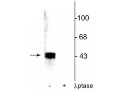
- Western blot of ERK1/2 in human T47D cell lysate showing specific immunolabeling of a ~42-44 kDa band corresponding to Phospho-ERK1/ERK2 (Thr202, Tyr204) polyclonal antibody (Product # 36-8800) in the first lane (-). Phosphospecificity is shown in the second lane (+) where immunolabeling is completely eliminated by blot treatment with lambda phosphatase (1,200 units for 30 min).

- Submitted by
- Invitrogen Antibodies (provider)
- Main image

- Experimental details
- Western blot analysis of p44 MAPK + p42 MAPK (pT202 + pT204) was performed by loading 30 µg of A431 (lane1), A431 treated for 10 minutes with 50 ng/mL PDGF (lane2), HeLa (lane3), HeLa treated for 10 minutes with 1:250 dilution of PDGF (lane4), NIH/3T3 (lane5), PC-12 (lane6), A549 (lane7), lysate using Novex® NuPAGE® 4-12 % Bis-Tris gel (Product # NP0322BOX), XCell SureLock™ Electrophoresis System (Product # EI0002), Novex® Sharp Pre-Stained Protein Standard (LC5800), and iBlot® Dry Blotting System (IB21001). Proteins were transferred to a nitrocellulose membrane and blocked with 5 % skim milk for 1 hour at room temperature. p44 MAPK + p42 MAPK (pT202 + pT204) was detected at 42, 44 kDa using p44 MAPK + p42 MAPK (pT202 + pT204) Rabbit Polyclonal Antibody (Product # 36-8800) at 1- 3 µg/mL in 5 % skim milk at 4°C overnight on a rocking platform. Goat Anti-Rabbit IgG - HRP Secondary Antibody (G21234) at 1:5000 dilution was used and chemiluminescent detection was performed using Pierce™ ECL Western Blotting Substrate (Product # 32106).

Supportive validation
- Submitted by
- Invitrogen Antibodies (provider)
- Main image

- Experimental details
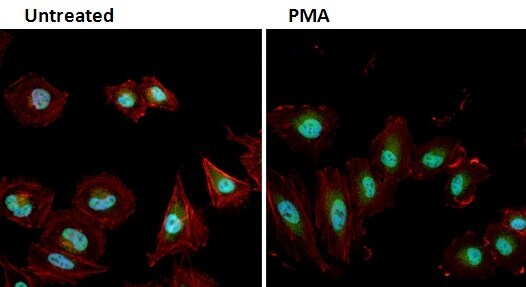
- Immunofluorescent analysis of Phospho-MAPK1/3 pThr202/Tyr204 (green) in HeLa cells either left untreated (left panel) or treated with 50nM PMA (right panel) for 10 minutes. Formalin fixed cells were permeabilized with 0.1% Triton X-100 in TBS for 10 minutes at room temperature and blocked with 1% Blocker BSA (Product # 37525) for 15 minutes at room temperature. Cells were probed with a Phospho-MAPK1/3 pThr202/Tyr204 polyclonal antibody (Product # 36-8800) at a dilution of 1:100 for at least 1 hour at room temperature, washed with PBS, and incubated with DyLight 488 goat anti-rabbit IgG secondary antibody (Product # 35552) at a dilution of 1:400 for 30 minutes at room temperature. F-Actin (red) was stained with DyLight 554 Phalloidin (Product # 21834) and nuclei (blue) were stained with Hoechst 33342 dye (Product # 62249). Images were taken on a Thermo Scientific ArrayScan or ToxInsight Instrument at 20X magnification.
- Submitted by
- Invitrogen Antibodies (provider)
- Main image
- Experimental details
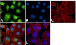
- Immunofluorescent analysis of Phospho-p44 MAPK + p42 MAPK pThr202 + pTyr204 Antibody was done on 70% confluent log phase A549 cells. The cells were fixed with 4% paraformaldehyde for 15 minutes, permeabilized with 0.25% Triton™ X-100 for 10 minutes, and blocked with 5% BSA for 1 hour at room temperature. The cells were labeled with Phospho-p44 MAPK + p42 MAPK pThr202 + pTyr204 Antibody (Product # 36-8800) at 1:250 dilution in 1% BSA and incubated for 3 hours at room temperature and then labeled with Alexa Fluor 488 Goat Anti-Rabbit IgG Secondary Antibody (Product # A-11008) at a dilution of 1:400 for 45 minutes at room temperature (Panel a: green). Nuclei (Panel b: blue) were stained with SlowFade® Gold Antifade Mountant with DAPI (Product # S36938). F-actin (Panel c: red) was stained with Alexa Fluor 594 Phalloidin (Product # A12381). Panel d is a merged image showing cytoplasmic and nuclear localization. Panel e is a no primary antibody control. The images were captured at 40X magnification.

Supportive validation
- Submitted by
- Invitrogen Antibodies (provider)
- Main image

- Experimental details
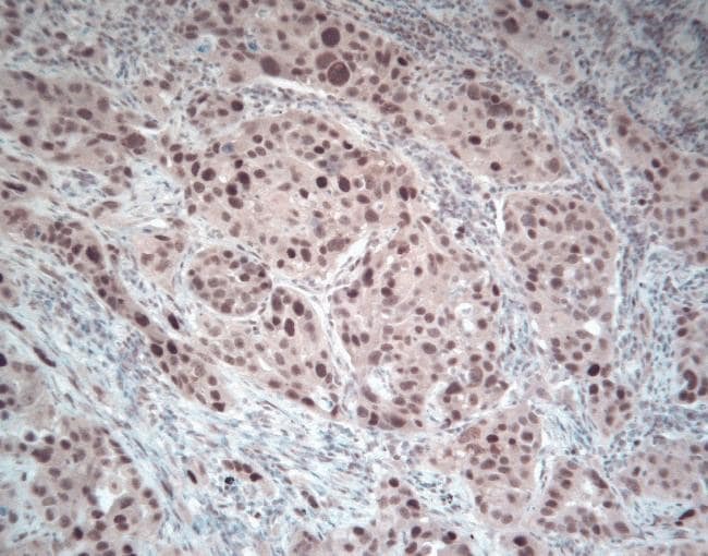
- Immunohistochemical staining of paraffin-embedded human lung cancer tissue using Zymed Rb anti-phospho-ERK1+2 (Thr202/Tyr204) (Product # 36-8800).
Supportive validation
- Submitted by
- Invitrogen Antibodies (provider)
- Main image

- Experimental details
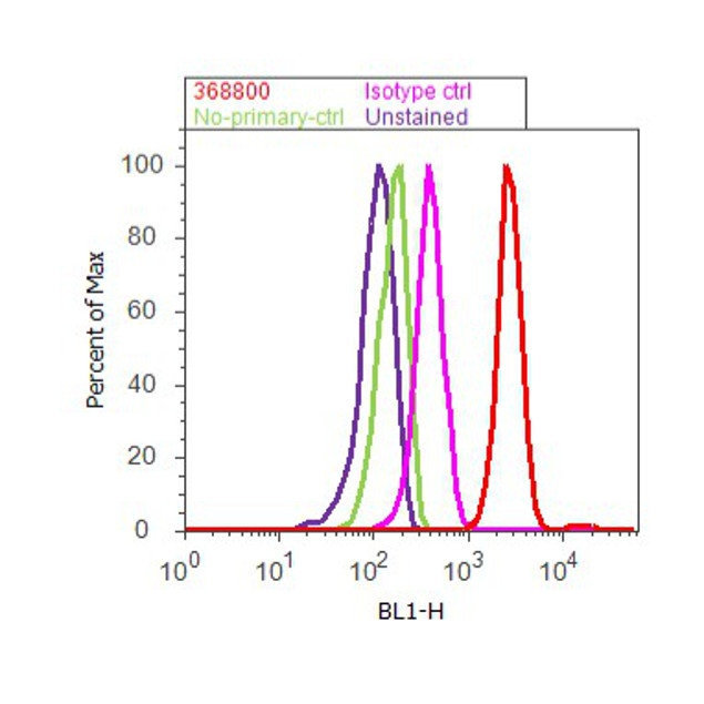
- Flow cytometry analysis of p44 MAPK+ p42 MAPK [pT202+ pY204] was done on K562 cells treated with TPA (200nM, 20 minutes). Cells were fixed with 70% ethanol for 10 minutes, permeabilized with 0.25% Triton™ X-100 for 20 minutes, and blocked with 5% BSA for 30 minutes at room temperature. Cells were labeled with p44 MAPK+ p42 MAPK [pT202+ pY204] Rabbit Polyclonal Antibody (368800, red histogram) or with rabbit isotype control (pink histogram) at 3-5 ug/million cells in 2.5% BSA. After incubation at room temperature for 2 hours, the cells were labeled with Alexa Fluor® 488 Goat Anti-Rabbit Secondary Antibody (A11008) at a dilution of 1:400 for 30 minutes at room temperature. The representative 10,000 cells were acquired and analyzed for each sample using an Attune® Acoustic Focusing Cytometer. The purple histogram represents unstained control cells and the green histogram represents no-primary-antibody control.
Supportive validation
- Submitted by
- Invitrogen Antibodies (provider)
- Main image

- Experimental details
- NULL
- Submitted by
- Invitrogen Antibodies (provider)
- Main image
- Experimental details
- NULL

- Submitted by
- Invitrogen Antibodies (provider)
- Main image

- Experimental details
- NULL
- Submitted by
- Invitrogen Antibodies (provider)
- Main image

- Experimental details
- NULL

- Submitted by
- Invitrogen Antibodies (provider)
- Main image
- Experimental details
- NULL

- Submitted by
- Invitrogen Antibodies (provider)
- Main image
- Experimental details
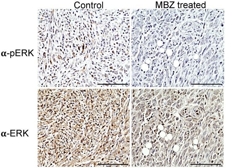
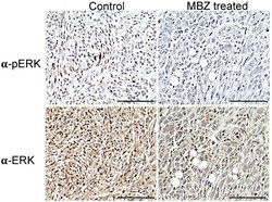
- Figure 4 MBZ reduces ERK (pERK) in treated NPcis mice. Representative images of tumors from untreated controls (left) and MBZ-treated NPcis mice (left) were stained for pERK1/2 (upper row) and ERK1/2 (lower row). pERK staining was visualized in brown in untreated controls but reduced in tumors of MBZ-treated mice. Each scale bar represents 100 um.