Explore
 Validate
 Learn
LearnPA5-14088
antibody from Invitrogen Antibodies
Targeting: MAPK1
ERK, ERK2, MAPK2, p41mapk, PRKM1, PRKM2
 Western blot
 Immunohistochemistry
Antibody data
- Antibody Data
- Antigen structure
- References [0]
- Comments [0]
- Validations
- Immunohistochemistry [1]
- Flow cytometry [2]
Submit
Validation data
Reference
Comment
Report error
- Product number
- PA5-14088 - Provider product page

- Provider
- Invitrogen Antibodies
- Product name
- ERK2 Polyclonal Antibody
- Antibody type
- Polyclonal
- Antigen
- Synthetic peptide
- Description
- This antibody is predicted to react with bovine, mouse, rat and Xenopus based on sequence homology.
- Reactivity
- Human
- Host
- Rabbit
- Isotype
- IgG
- Vial size
- 400 μL
- Concentration
- 2 mg/mL
- Storage
- Store at 4°C short term. For long term storage, store at -20°C, avoiding freeze/thaw cycles.
No comments: Submit comment
Supportive validation
- Submitted by
- Invitrogen Antibodies (provider)
- Main image
- Experimental details
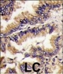
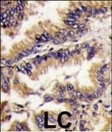
- Immunohistochemistry analysis of ERK2 in formalin-fixed and paraffin-embedded human lung carcinoma tissue. Samples were incubated with ERK2 polyclonal antibody (Product # PA5-14088) which was peroxidase-conjugated to the secondary antibody, followed by DAB staining. This data demonstrates the use of this antibody for immunohistochemistry; clinical relevance has not been evaluated.
Supportive validation
- Submitted by
- Invitrogen Antibodies (provider)
- Main image

- Experimental details
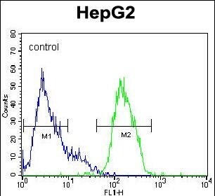
- Flow cytometry analysis of HepG2 cells using an ERK2 polyclonal antibody (Product # PA5-14088) (right) compared to a negative control cell (left) at a dilution of 1:10-50, followed by a FITC-conjugated goat anti-rabbit antibody
- Submitted by
- Invitrogen Antibodies (provider)
- Main image
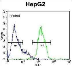
- Experimental details
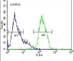
- Flow cytometry of ERK2 in HepG2 cells (right histogram). Samples were incubated with ERK2 polyclonal antibody (Product # PA5-14088) followed by FITC-conjugated goat-anti-rabbit secondary antibody. Negative control cell (left histogram).