 Explore
 Validate
 Learn
LearnPA5-29636
antibody from Invitrogen Antibodies
Targeting: MAPK1
ERK, ERK2, MAPK2, p41mapk, PRKM1, PRKM2
 Western blot
 Immunoprecipitation
Antibody data
- Antibody Data
- Antigen structure
- References [2]
- Comments [0]
- Validations
- Immunoprecipitation [1]
- Immunohistochemistry [2]
- Other assay [3]
Submit
Validation data
Reference
Comment
Report error
- Product number
- PA5-29636 - Provider product page

- Provider
- Invitrogen Antibodies
- Product name
- ERK2 Polyclonal Antibody
- Antibody type
- Polyclonal
- Antigen
- Recombinant full-length protein
- Description
- Recommended positive controls: 293T, A431, H1299, HeLa, HepG2, Molt-4, Raji, mouse brain, SD Rat brain thalamus, SD Rat brain amygdala. Predicted reactivity: Mouse (100%), Rat (100%), Zebrafish (96%), Xenopus laevis (97%), Dog (100%), Pig (99%), Chicken (99%), Bovine (99%). Store product as a concentrated solution. Centrifuge briefly prior to opening the vial.
- Reactivity
- Human, Mouse, Rat, Porcine
- Host
- Rabbit
- Isotype
- IgG
- Vial size
- 100 μL
- Concentration
- 1.0 mg/mL
- Storage
- Store at 4°C short term. For long term storage, store at -20°C, avoiding freeze/thaw cycles.
Submitted references OGDH is involved in sepsis induced acute lung injury through the MAPK pathway.
Curcumin Enhances Radiosensitization of Nasopharyngeal Carcinoma via Mediating Regulation of Tumor Stem-like Cells by a CircRNA Network.
Hao Y, Wang Z, Wang X, Zhan W, Wu D
Journal of thoracic disease 2021 Aug;13(8):5042-5054
Journal of thoracic disease 2021 Aug;13(8):5042-5054
Curcumin Enhances Radiosensitization of Nasopharyngeal Carcinoma via Mediating Regulation of Tumor Stem-like Cells by a CircRNA Network.
Zhu D, Shao M, Yang J, Fang M, Liu S, Lou D, Gao R, Liu Y, Li A, Lv Y, Mo Z, Fan Q
Journal of Cancer 2020;11(8):2360-2370
Journal of Cancer 2020;11(8):2360-2370
No comments: Submit comment
Supportive validation
- Submitted by
- Invitrogen Antibodies (provider)
- Main image

- Experimental details
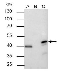
- ERK2 Polyclonal Antibody immunoprecipitates MAPK1 protein in IP experiments. IP samples: HeLa whole cell extract. A. 30 µg HeLa whole cell extract. B. Control with 4 µg of preimmune Rabbit IgG. C. Immunoprecipitation of MAPK1 protein by 4 µg ERK2 Polyclonal Antibody (Product # PA5-29636). 10 % SDS-PAGE. The immunoprecipitated MAPK1 protein was detected by ERK2 Polyclonal Antibody (Product # PA5-29636) diluted at 1:500.

Supportive validation
- Submitted by
- Invitrogen Antibodies (provider)
- Main image
- Experimental details
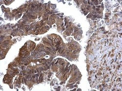
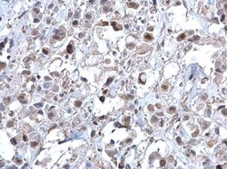
- ERK2 Polyclonal Antibody detects ERK2 protein at cytosol and nucleus on human colon carcinoma by immunohistochemical analysis. Sample: Paraffin-embedded human colon carcinoma. ERK2 Polyclonal Antibody (Product # PA5-29636) dilution: 1:500. Antigen Retrieval: EDTA based buffer, pH 8.0, 15 min.
- Submitted by
- Invitrogen Antibodies (provider)
- Main image
- Experimental details
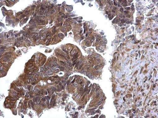
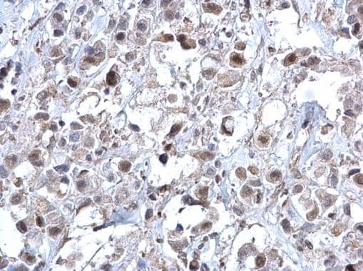
- ERK2 Polyclonal Antibody detects ERK2 protein at cytosol and nucleus on human breast carcinoma by immunohistochemical analysis. Sample: Paraffin-embedded human breast carcinoma. ERK2 Polyclonal Antibody (Product # PA5-29636) dilution: 1:500. Antigen Retrieval: EDTA based buffer, pH 8.0, 15 min.
Supportive validation
- Submitted by
- Invitrogen Antibodies (provider)
- Main image
- Experimental details
- ERK2 Polyclonal Antibody immunoprecipitates MAPK1 protein in IP experiments. IP samples: HeLa whole cell extract. A. 30 µg HeLa whole cell extract. B. Control with 4 µg of preimmune Rabbit IgG. C. Immunoprecipitation of MAPK1 protein by 4 µg ERK2 Polyclonal Antibody (Product # PA5-29636). 10 % SDS-PAGE. The immunoprecipitated MAPK1 protein was detected by ERK2 Polyclonal Antibody (Product # PA5-29636) diluted at 1:500.

- Submitted by
- Invitrogen Antibodies (provider)
- Main image
- Experimental details
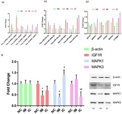
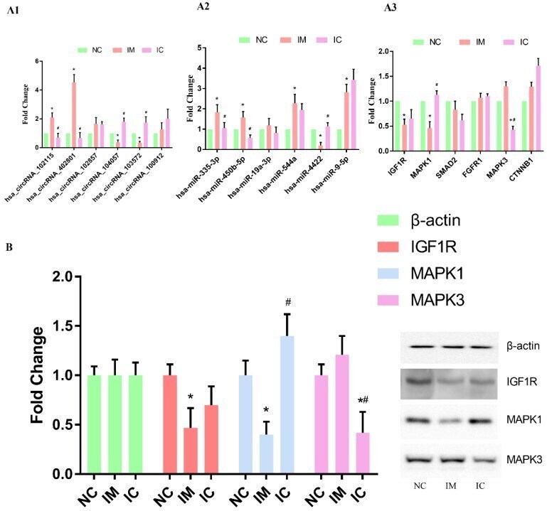
- Figure 4 Verification of the circRNA-miRNA-mRNA interaction network. (A1) qRT-PCR detection of circRNA expression in the three groups. (A2) Levels of target miRNAs for circRNA in the three groups. (A3) Levels of target mRNAs for miRNAs in the three groups. The expression of each RNA was calculated using 2-^^Ct equation. Compared to NC group: *p < 0.05, and compared to IM group: #p < 0.05. (B) WB analysis of selected protein expression. Compared to NC group: *p < 0.05, and compared to IM group: #p < 0.05.
- Submitted by
- Invitrogen Antibodies (provider)
- Main image
- Experimental details
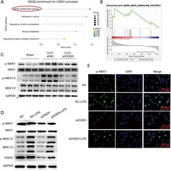
- Figure 5 MAPK is the downstream pathway of OGDH. (A) According to the expression data of GSE16650, the KEGG analysis was used to generate the bubble plot; (B) GSEA analysis showed that the gene set 'KEGG_MAPK_SIGNALING_pathway' was highly expressed in the high expression group of OGDH; (C) in the CLP animal model, down-regulation of OGDH reversed the increase of p-MAPK1 expression; (D) in an LPS-induced lung injury cell model, down-regulation of OGDH can down-regulate p-MAPK1 expression; (E) immunofluorescence showed down-regulation of OGDH and down-regulation of p-MAPK1 expression. OGDH, oxoglutarate dehydrogenase.
