Explore
 Validate
 Learn
LearnPA5-32396
antibody from Invitrogen Antibodies
Targeting: MAPK1
ERK, ERK2, MAPK2, p41mapk, PRKM1, PRKM2
 Western blot
 Immunohistochemistry
Antibody data
- Antibody Data
- Antigen structure
- References [0]
- Comments [0]
- Validations
- Western blot [2]
Submit
Validation data
Reference
Comment
Report error
- Product number
- PA5-32396 - Provider product page

- Provider
- Invitrogen Antibodies
- Product name
- Anti-ERK2 Polyclonal Antibody
- Antibody type
- Polyclonal
- Antigen
- Synthetic peptide
- Description
- This antibody is predicted to react with bovine, chicken, canine, amphibian, porcine, rabbit and rat based on sequence homology. Heat-mediated antigen retrieval is recommended prior to staining, using a 10mM citrate buffer, pH 6.0, for 10 minutes followed by cooling at room temperature for 20 min. Following antigen retrieval, incubate samples with primary antibody for 10 min at room temperature. A suggested positive control is breast carcinoma.
- Reactivity
- Human, Mouse
- Host
- Rabbit
- Isotype
- IgG
- Vial size
- 500 µL
- Storage
- -20° C, Avoid Freeze/Thaw Cycles
No comments: Submit comment
Supportive validation
- Submitted by
- Invitrogen Antibodies (provider)
- Main image
- Experimental details
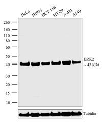
- Western blot analysis was performed on whole cell extracts (30 µg lysate) of HeLa (Lane 1), H1975 (Lane 2), HCT 116 (Lane 3), HT-29 (Lane 4), A-431 (Lane 5) and A549 (Lane 6). The blots were probed with Anti-ERK2 Rabbit Polyclonal Antibody (Product # PA5-32396, 1:1000 dilution) and detected by chemiluminescence using Goat anti-Rabbit IgG (H+L) Superclonal™ Secondary Antibody, HRP conjugate (Product # A27036, 0.25 µg/mL, 1:4000 dilution). A 42 kDa band corresponding to ERK2 was observed across the cell lines tested. Known quantity of protein samples were electrophoresed using Novex® NuPAGE® 4-12 % Bis-Tris gel (Product # NP0321BOX), XCell SureLock™ Electrophoresis System (Product # EI0002) and Novex® Sharp Pre-Stained Protein Standard (Product # LC5800). Resolved proteins were then transferred onto a nitrocellulose membrane with iBlot® 2 Dry Blotting System (Product # IB21001). The membrane was probed with the relevant primary and secondary Antibody following blocking with 5 % skimmed milk. Chemiluminescent detection was performed using Pierce™ ECL Western Blotting Substrate (Product # 32106).

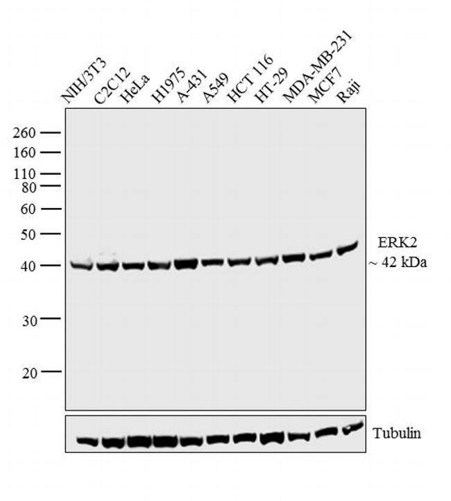
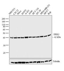
- Submitted by
- Invitrogen Antibodies (provider)
- Main image
- Experimental details
- Western blot analysis was performed on whole cell extracts (30 µg lysate) of NIH/3T3 (Lane 1), C2C12 (Lane 2), HeLa (Lane 3), H1975 (Lane 4), A-431 (Lane 5), A549 (Lane 6), HCT 116 (Lane 7), HT-29 (Lane 8), MDA-MB-231 (Lane 9), MCF7 (Lane 10) and Raji (Lane 11). The blot was probed with Rabbit Anti-ERK2 Polyclonal Antibody (Product # PA1-37640, 1:1000 dilution) and detected by chemiluminescence using Goat anti-Rabbit IgG (H+L) Superclonal™ Secondary Antibody, HRP conjugate (Product # A27036, 0.25 µg/mL, 1:4000 dilution). A 42 kDa band corresponding to ERK2 was observed across the cell lines tested. Known quantity of protein samples were electrophoresed using Novex® NuPAGE® 10 % Bis-Tris gel (Product # NP0302BOX), XCell SureLock™ Electrophoresis System (Product # EI0002) and Novex® Sharp Pre-Stained Protein Standard (Product # LC5800). Resolved proteins were then transferred onto a nitrocellulose membrane with iBlot® 2 Dry Blotting System (Product # IB21001). The membrane was probed with the relevant primary and secondary Antibody following blocking with 5 % skimmed milk. Chemiluminescent detection was performed using Pierce™ ECL Western Blotting Substrate (Product # 32106).