Explore
 Validate
 Learn
Learn Western blot
Antibody data
- Antibody Data
- Antigen structure
- References [0]
- Comments [0]
- Validations
- Western blot [2]
- Immunohistochemistry [1]
Submit
Validation data
Reference
Comment
Report error
- Product number
- AF1230 - Provider product page

- Provider
- R&D Systems
- Product name
- Human/Mouse/Rat ERK2 Antibody
- Antibody type
- Polyclonal
- Description
- Antigen and protein A Affinity-purified. Detects human, mouse, and rat ERK2. Also recognizes ERK1 in some cell types at much lower affinity.
- Reactivity
- Human, Mouse, Rat
- Host
- Rabbit
- Conjugate
- Unconjugated
- Antigen sequence
P28482- Isotype
- IgG
- Vial size
- 100 ug
- Concentration
- LYOPH
- Storage
- Use a manual defrost freezer and avoid repeated freeze-thaw cycles. 12 months from date of receipt, -20 to -70 °C as supplied. 1 month, 2 to 8 °C under sterile conditions after reconstitution. 6 months, -20 to -70 °C under sterile conditions after reconstitution.
No comments: Submit comment
Supportive validation
- Submitted by
- R&D Systems (provider)
- Main image
- Experimental details
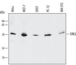
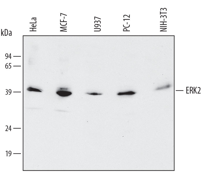
- Detection of Human/Mouse/Rat ERK2 by Western Blot. Western blot shows lysates of HeLa human cervical epithelial carcinoma cell line, MCF-7 human breast cancer cell line, U937 human histiocytic lymphoma cell line, PC-12 rat adrenal pheochromocytoma cell line, and NIH-3T3 mouse embryonic fibroblast cell line. PVDF membrane was probed with 0.1 µg/mL of Rabbit Anti-Human/Mouse/Rat ERK2 Antigen Affinity-purified Polyclonal Antibody (Catalog # AF1230) followed by HRP-conjugated Anti-Rabbit IgG Secondary Antibody (Catalog # HAF008). A specific band was detected for ERK2 at approximately 42 kDa (as indicated). This experiment was conducted under reducing conditions and using Immunoblot Buffer Group 1.
- Submitted by
- R&D Systems (provider)
- Main image

- Experimental details
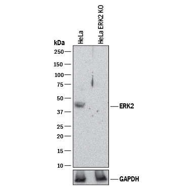
- Western Blot Shows Human ERK2 Specificity by Using Knockout Cell Line. Western blot shows lysates of HeLa human cervical epithelial carcinoma parental cell line and ERK2 knockout HeLa cell line (KO). PVDF membrane was probed with 0.1 µg/mL of Rabbit Anti-Human/Mouse/Rat ERK2 Antigen Affinity-purified Polyclonal Antibody (Catalog # AF1230) followed by HRP-conjugated Anti-Rabbit IgG Secondary Antibody (Catalog # HAF008). A specific band was detected for ERK2 at approximately 42 kDa (as indicated) in the parental HeLa cell line, but is not detectable in knockout HeLa cell line. GAPDH (Catalog # AF5718) is shown as a loading control. This experiment was conducted under reducing conditions and using Immunoblot Buffer Group 1.
Supportive validation
- Submitted by
- R&D Systems (provider)
- Main image
- Experimental details
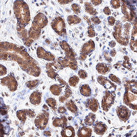
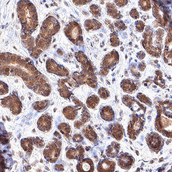
- ERK2 in Human Breast. ERK2 was detected in immersion fixed paraffin-embedded sections of human breast using Rabbit Anti-Human/Mouse/Rat ERK2 Antigen Affinity-purified Polyclonal Antibody (Catalog # AF1230) at 3 µg/mL for 1 hour at room temperature followed by incubation with the Anti-Rabbit IgG VisUCyte™ HRP Polymer Antibody (Catalog # VC003). Tissue was stained using DAB (brown) and counterstained with hematoxylin (blue). Specific staining was localized to cytoplasm in epithelial cells. View our protocol for IHC Staining with VisUCyte HRP Polymer Detection Reagents.