Explore
 Validate
 Learn
Learn Western blot
Antibody data
- Antibody Data
- Antigen structure
- References [3]
- Comments [0]
- Validations
- Western blot [2]
- Immunocytochemistry [1]
- Immunohistochemistry [4]
- Flow cytometry [2]
Submit
Validation data
Reference
Comment
Report error
- Product number
- GTX84377 - Provider product page

- Provider
- GeneTex
- Proper citation
- GeneTex Cat#GTX84377, RRID:AB_10728413
- Product name
- HDAC6 antibody [4C5]
- Antibody type
- Monoclonal
- Reactivity
- Human, Mouse, Rat, Canine, Simian
- Host
- Mouse
Submitted references Clinacanthus nutans Protects Cortical Neurons Against Hypoxia-Induced Toxicity by Downregulating HDAC1/6.
Primary cilia in stem cells and neural progenitors are regulated by neutral sphingomyelinase 2 and ceramide.
Characterization of an apical ceramide-enriched compartment regulating ciliogenesis.
Tsai HD, Wu JS, Kao MH, Chen JJ, Sun GY, Ong WY, Lin TN
Neuromolecular medicine 2016 Sep;18(3):274-82
Neuromolecular medicine 2016 Sep;18(3):274-82
Primary cilia in stem cells and neural progenitors are regulated by neutral sphingomyelinase 2 and ceramide.
He Q, Wang G, Wakade S, Dasgupta S, Dinkins M, Kong JN, Spassieva SD, Bieberich E
Molecular biology of the cell 2014 Jun;25(11):1715-29
Molecular biology of the cell 2014 Jun;25(11):1715-29
Characterization of an apical ceramide-enriched compartment regulating ciliogenesis.
He Q, Wang G, Dasgupta S, Dinkins M, Zhu G, Bieberich E
Molecular biology of the cell 2012 Aug;23(16):3156-66
Molecular biology of the cell 2012 Aug;23(16):3156-66
No comments: Submit comment
Supportive validation
- Submitted by
- GeneTex (provider)
- Main image
- Experimental details
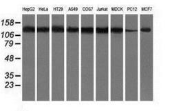
- Western blot analysis of extracts (35ug) from 9 different cell lines by using anti-HDAC6 monoclonal antibody.

- Submitted by
- GeneTex (provider)
- Main image

- Experimental details
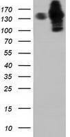
- HEK293T cells were transfected with the pCMV6-ENTRY control (Left lane) or pCMV6-ENTRY HDAC6 (Right lane) cDNA for 48 hrs and lysed. Equivalent amounts of cell lysates (5 ug per lane) were separated by SDS-PAGE and immunoblotted with anti-HDAC6.
Supportive validation
- Submitted by
- GeneTex (provider)
- Main image

- Experimental details
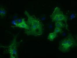
- Anti-HDAC6 mouse monoclonal antibody (GTX84377) immunofluorescent staining of COS7 cells transiently transfected with HDAC6
Supportive validation
- Submitted by
- GeneTex (provider)
- Main image
- Experimental details
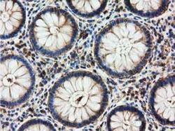
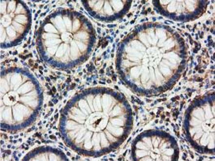
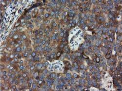
- IHC-P analysis of human colon tissue using GTX84377 HDAC6 antibody [4C5].Dilution : 1:150
- Submitted by
- GeneTex (provider)
- Main image
- Experimental details
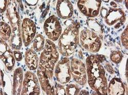
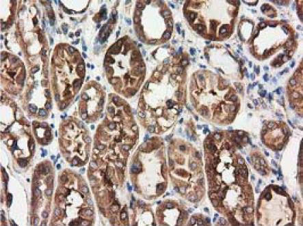
- IHC-P analysis of human kidney tissue using GTX84377 HDAC6 antibody [4C5].Dilution : 1:150
- Submitted by
- GeneTex (provider)
- Main image

- Experimental details
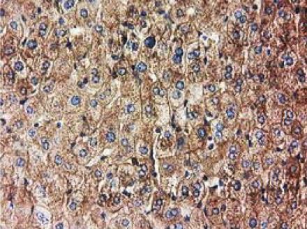
- IHC-P analysis of human liver tissue using GTX84377 HDAC6 antibody [4C5].Dilution : 1:150
- Submitted by
- GeneTex (provider)
- Main image
- Experimental details
- IHC-P analysis of human ovary tissue using GTX84377 HDAC6 antibody [4C5].Dilution : 1:150

Supportive validation
- Submitted by
- GeneTex (provider)
- Main image
- Experimental details
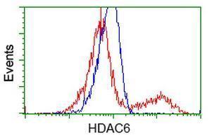
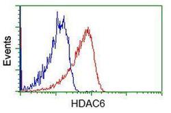
- Flow cytometric Analysis of Jurkat cells, using anti-HDAC6 antibody(GTX84377),(Red), compared to a nonspecific negative control antibody(TA50011),(Blue).
- Submitted by
- GeneTex (provider)
- Main image

- Experimental details
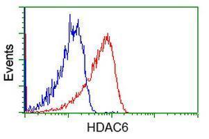
- HEK293T cells transfected with either RC209649 overexpress plasmid(Red) or empty vector control plasmid(Blue) were immunostained by anti-HDAC6 antibody(GTX84377), and then analyzed by flow cytometry.