Explore
 Validate
 Learn
LearnMA5-60183
antibody from Invitrogen Antibodies
Targeting: LAMC2
BM600-100kDa, EBR2, EBR2A, kalinin-105kDa, LAMB2T, LAMNB2, nicein-100kDa
 Western blot
 Immunocytochemistry
Antibody data
- Antibody Data
- Antigen structure
- References [0]
- Comments [0]
- Validations
- Immunocytochemistry [3]
- Immunohistochemistry [5]
- Flow cytometry [1]
Submit
Validation data
Reference
Comment
Report error
- Product number
- MA5-60183 - Provider product page

- Provider
- Invitrogen Antibodies
- Product name
- Laminin gamma-2 Recombinant Rabbit Monoclonal Antibody (PSH07-27)
- Antibody type
- Monoclonal
- Antigen
- Recombinant full-length protein
- Description
- Positive Control: A431 cell lysate, A549 cell lysate, HaCaT cell lysate, NCI-H441 cell lysate, Mouse skin tissue lysate, Mouse stomach tissue lysate, Rat skin tissue lysate, Rat stomach tissue lysate, human colon tissue, mouse colon tissue, rat colon tissue, A431. Tissue Specificity: Tissue enhanced (urinary). Subcellular Location: Secreted, extracellular space, extracellular matrix, basement membrane. Sequence Similarities: 78% Mouse/Rat. Predicted band size: 131 kDa.
- Reactivity
- Human, Mouse, Rat
- Host
- Rabbit
- Isotype
- IgG
- Antibody clone number
- PSH07-27
- Vial size
- 100 μL
- Concentration
- 1 mg/mL
- Storage
- Store at 4°C short term. For long term storage, store at -20°C, avoiding freeze/thaw cycles.
No comments: Submit comment
Supportive validation
- Submitted by
- Invitrogen Antibodies (provider)
- Main image

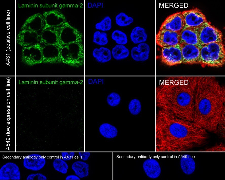
- Experimental details
- Immunofluorescence analysis of LAMC2 using A431 cells (positive) (top) and A549 cells (low expression) (bottom).The cells were fixed in 4% paraformaldehyde for 20 minutes at room temperature, permeabilized with 0.1% Triton X-100 in PBS for 5 minutes at room temperature, then blocked with 1% BSA in 10% negative goat serum for 1 hour at room temperature. The cells were co-labeled with Laminin gamma-2 Recombinant Rabbit Monoclonal Antibody (PSH07-27) (Product # MA5-60183, green) and beta tubulin (red) at 1:100 dilution in 1% BSA in PBST overnight at 4 degrees Celsius and then labeled with iFluor™ 488 Goat Anti-Rabbit IgG H&L and iFluor™ 594 Anti-Mouse IgG H&L secondary antibodies for 1 hour at room temperature. PBS instead of the primary antibody was used as the secondary antibody only control. Nuclei were stained with DAPI (blue). The images were captured at 200X magnification.
- Submitted by
- Invitrogen Antibodies (provider)
- Main image
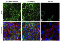
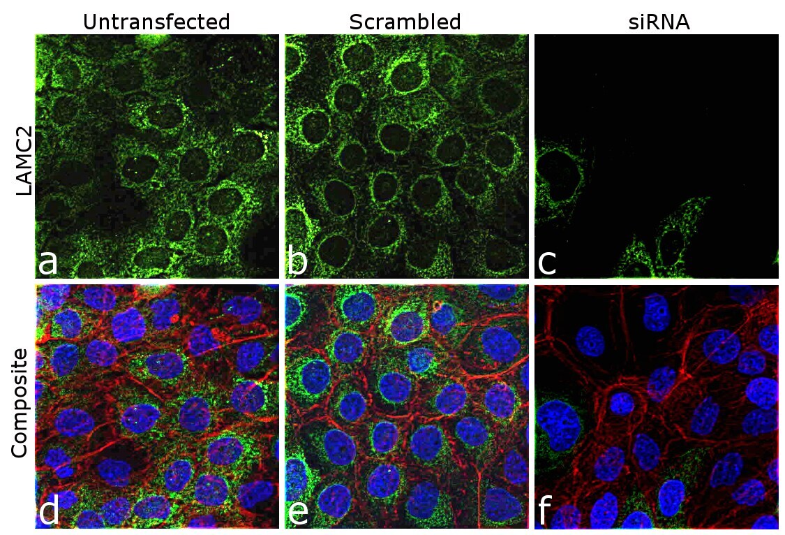
- Experimental details
- Knockdown of Laminin gamma-2 (LAMC2) was achieved by transfecting A-431 cells with specific siRNA (Silencer® select Product # s8084, s534191). Immunofluorescence analysis was performed on A-431 cells (untransfected, panels a,d), transfected with LAMC2 specific siRNA (panels c,f) or non-specific scrambled siRNA (panels b,e). Cells were fixed, permeabilized, and labeled with Laminin gamma-2 Recombinant Rabbit Monoclonal Antibody (PSH07-27) (Product # MA5-60183, 1:100 dilution), followed by Goat anti-Rabbit IgG (H+L) Highly Cross-Adsorbed Secondary Antibody, Alexa Fluor™ Plus 488 (Product # A32731, 1:2,000 dilution). Nuclei (blue) were stained using ProLong™ Diamond Antifade Mountant with DAPI (Product # P36962), and Rhodamine Phalloidin (Product # R415, 1:300 dilution) was used for cytoskeletal F-actin (red) staining. Significant reduction of signal was observed upon siRNA mediated knockdown (panel c,f) confirming specificity of the antibody to LAMC2 (green). The images were captured at 40X magnification.
- Submitted by
- Invitrogen Antibodies (provider)
- Main image
- Experimental details
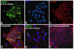
- Immunofluorescence analysis of Laminin gamma-2 (LAMC2) was performed using 70% confluent log phase A-431 and SH-SY5Y cells. The cells were fixed with 4% paraformaldehyde for 10 minutes, permeabilized with 0.1% Triton™ X-100 for 15 minutes, and blocked with 2% BSA for 1 hour at room temperature. The cells were labeled with Laminin gamma-2 Recombinant Rabbit Monoclonal Antibody (PSH07-27) (Product # MA5-60183) at 1:100 dilution in 0.1% BSA, incubated at 4 degree celsius overnight and then labeled with Goat anti-Rabbit IgG (H+L) Highly Cross-Adsorbed Secondary Antibody, Alexa Fluor™ Plus 488 (Product # A32731, 1:2,000), for 45 minutes at room temperature (Panel a: Green). Nuclei (Panel b:Blue) were stained with ProLong™ Diamond Antifade Mountant with DAPI (Product # P36962). F-actin (Panel c: Red) was stained with Rhodamine Phalloidin (Product # R415, 1:300). Panel d represents the merged image of A-431, which is a high expressing model for LAMC2 showing cytoplasmic localization. Panel e represents the merged image of SH-SY5Y cells that are a low expressing model for LAMC2. Panel f represents control cells A-431 with no primary antibody to assess background. The images were captured at 40x magnification.
Supportive validation
- Submitted by
- Invitrogen Antibodies (provider)
- Main image
- Experimental details
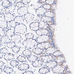
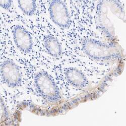
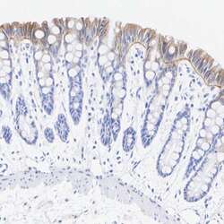
- Immunohistochemical analysis of LAMC2 on formalin-fixed paraffin-embedded mouse colon tissue.The section was pre-treated using heat mediated antigen retrieval with Tris-EDTA buffer (pH 9.0) for 20 minutes. The tissue was blocked with 1% BSA for 20 minutes at room temperature, then probed with Laminin gamma-2 Recombinant Rabbit Monoclonal Antibody (PSH07-27) (Product # MA5-60183) at 1:200 dilution for 1 hour at room temperature. HRP conjugated compact polymer system and DAB chromogen were used as the detection system, followed by counterstaining with hematoxylin. The slide was mounted with DPX and the image was captured at 200X magnification.
- Submitted by
- Invitrogen Antibodies (provider)
- Main image
- Experimental details
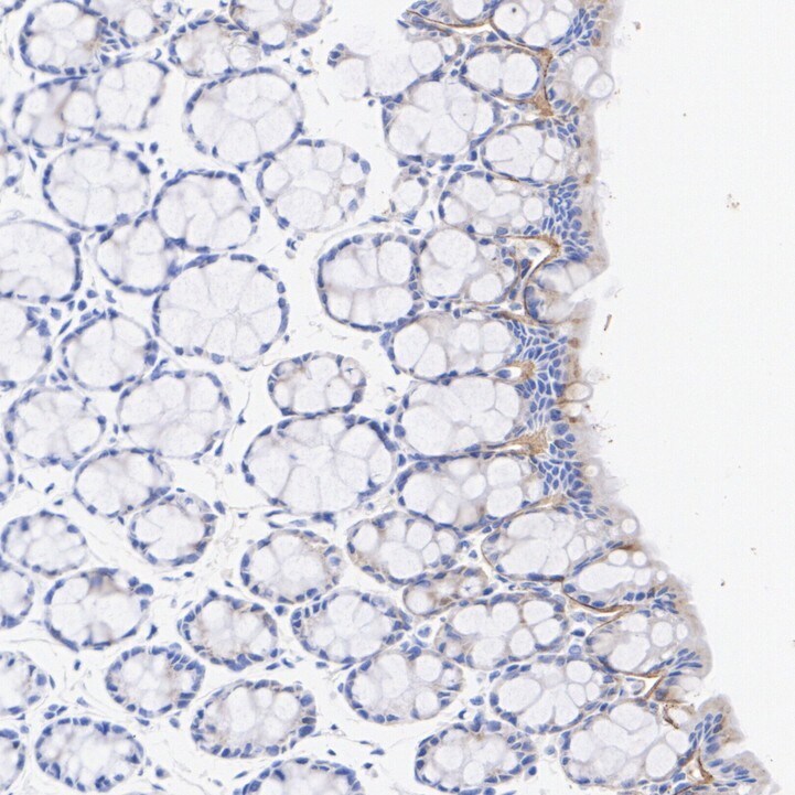
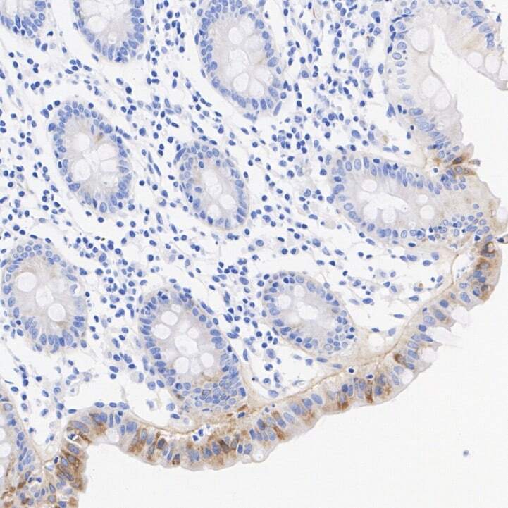
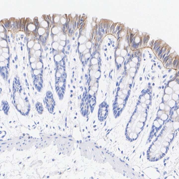
- Immunohistochemical analysis of LAMC2 on formalin-fixed paraffin-embedded human colon tissue.The section was pre-treated using heat mediated antigen retrieval with Tris-EDTA buffer (pH 9.0) for 20 minutes. The tissue was blocked with 1% BSA for 20 minutes at room temperature, then probed with Laminin gamma-2 Recombinant Rabbit Monoclonal Antibody (PSH07-27) (Product # MA5-60183) at 1:200 dilution for 1 hour at room temperature. HRP conjugated compact polymer system and DAB chromogen were used as the detection system, followed by counterstaining with hematoxylin. The slide was mounted with DPX and the image was captured at 200X magnification.
- Submitted by
- Invitrogen Antibodies (provider)
- Main image
- Experimental details
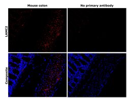
- Immunohistochemical analysis of Laminin gamma-2 (LAMC2) was performed using formalin-fixed paraffin-embedded mouse colon tissue sections. To expose the target protein, heat-induced epitope retrieval was performed on de-paraffinized sections using eBioscience™ IHC Antigen Retrieval Solution - High pH (10X) (Product # 00-4956-58) diluted to 1X solution in water in a decloaking chamber at 110 degree Celsius for 15 minutes. Following antigen retrieval, the sections were blocked with 3% H2O2 for 1 hour at room temperature followed by 2% Normal goat serum in 1X PBS for 45 minutes at room temperature and then probed with or without Laminin gamma-2 Recombinant Rabbit Monoclonal Antibody (PSH07-27) (Product # MA5-60183) at 1:500 dilution in 0.1% Normal goat serum overnight at 4 degree Celsius in a humidified chamber. Detection was performed using Alexa Fluor™ 594 Tyramide SuperBoost™ Kit, goat anti-rabbit IgG (Product # B40925). Nuclei were stained with DAPI (Product # D1306) and the sections were mounted using ProLong™ Glass Antifade Mountant (Product # P36984). The images were captured on EVOS™ M7000 Imaging System (Product # AMF7000) at 20X magnification and externally deconvoluted.
- Submitted by
- Invitrogen Antibodies (provider)
- Main image
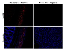
- Experimental details
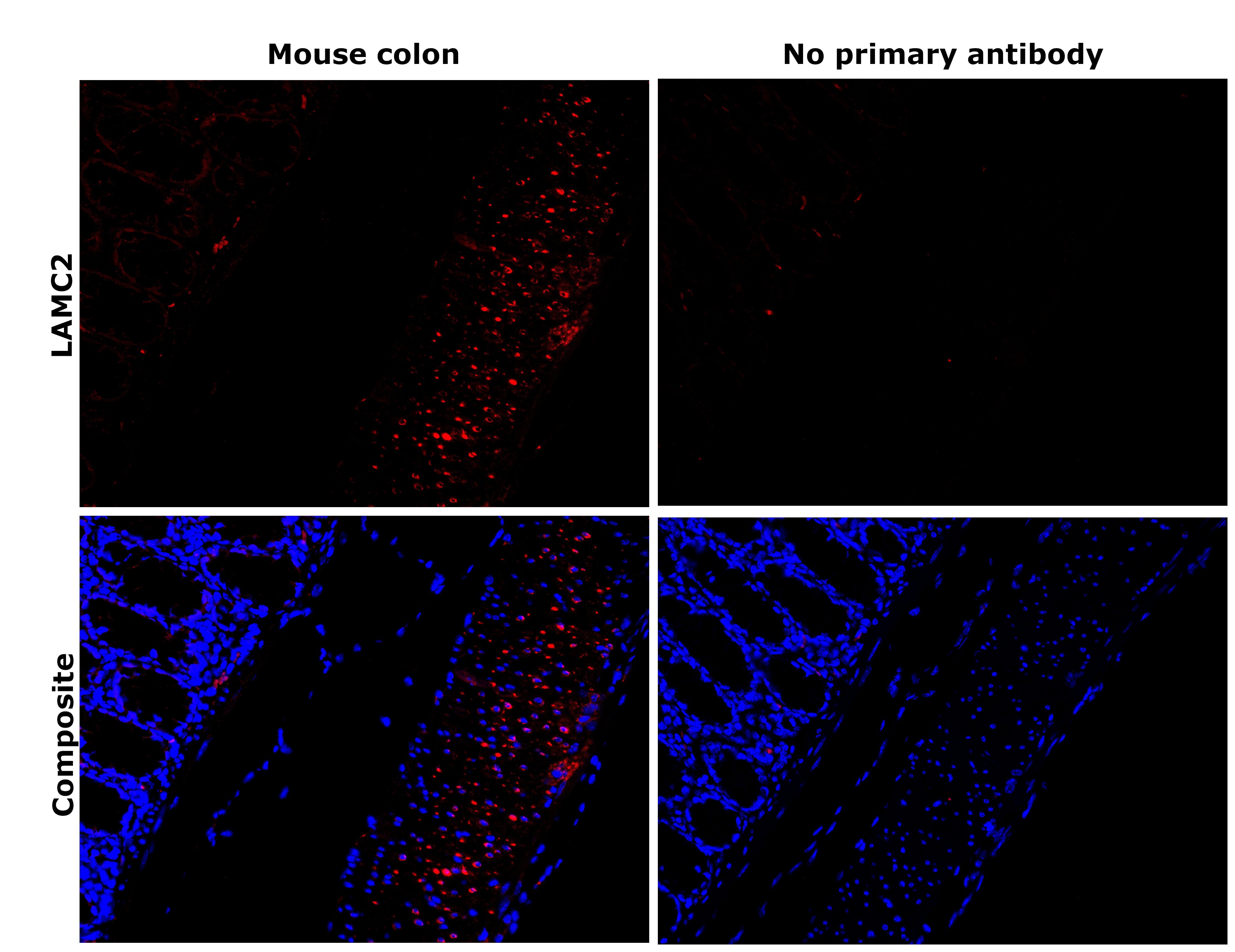
- Immunohistochemical analysis of Laminin gamma-2 (LAMC2) was performed using formalin-fixed paraffin-embedded mouse colon and mouse liver tissue sections. To expose the target protein, heat-induced epitope retrieval was performed on de-paraffinized sections using eBioscience™ IHC Antigen Retrieval Solution - High pH (10X) (Product # 00-4956-58) diluted to 1X solution in water in a decloaking chamber at 110 degree Celsius for 15 minutes. Following antigen retrieval, the sections were blocked with 3% H2O2 for 1 hour at room temperature followed by 2% Normal goat serum in 1X PBS for 45 minutes at room temperature and then probed with or without Laminin gamma-2 Recombinant Rabbit Monoclonal Antibody (PSH07-27) (Product # MA5-60183) at 1:500 dilution in 0.1% Normal goat serum overnight at 4 degree Celsius in a humidified chamber. Detection was performed using Alexa Fluor™ 594 Tyramide SuperBoost™ Kit, goat anti-rabbit IgG (Product # B40925). Nuclei were stained with DAPI (Product # D1306) and the sections were mounted using ProLong™ Glass Antifade Mountant (Product # P36984). The images were captured on EVOS™ M7000 Imaging System (Product # AMF7000) at 20X magnification and externally deconvoluted. A cytosolic localisation specific to LAMC2 was observed in the mucosal layer of the mouse colon tissue. No staining was observed in mouse liver tissue sections, which is known to express lower levels of LAMC2.
- Submitted by
- Invitrogen Antibodies (provider)
- Main image
- Experimental details
- Immunohistochemical analysis of LAMC2 on formalin-fixed paraffin-embedded rat colon tissue.The section was pre-treated using heat mediated antigen retrieval with Tris-EDTA buffer (pH 9.0) for 20 minutes. The tissue was blocked with 1% BSA for 20 minutes at room temperature, then probed with Laminin gamma-2 Recombinant Rabbit Monoclonal Antibody (PSH07-27) (Product # MA5-60183) at 1:200 dilution for 1 hour at room temperature. HRP conjugated compact polymer system and DAB chromogen were used as the detection system, followed by counterstaining with hematoxylin. The slide was mounted with DPX and the image was captured at 200X magnification.
Supportive validation
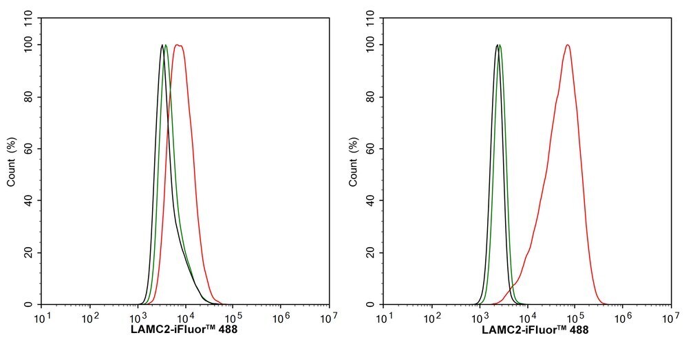
- Submitted by
- Invitrogen Antibodies (provider)
- Main image
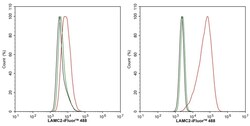
- Experimental details
- A431 cells (positive) (right) and A549 cells (low expression) (left) were fixed and permeabilized, and then stained with Laminin gamma-2 Recombinant Rabbit Monoclonal Antibody (PSH07-27) (Product # MA5-60183) (red) or with Rabbit IgG Isotype Control (green). After incubation of the primary antibody at 4 degrees Celsius for an hour, the cells were stained with a iFluor™ 488 conjugate-Goat anti-Rabbit IgG Secondary antibody at 1:1,000 dilution for 30 minutes at 4 degrees Celsius. Unlabelled sample was used as a control (cells without incubation with primary antibody; black).