Explore
 Validate
 Learn
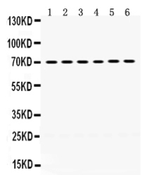
Learn Western blot
 Immunohistochemistry
Antibody data
- Antibody Data
- Antigen structure
- References [0]
- Comments [0]
- Validations
- Immunohistochemistry [1]
Submit
Validation data
Reference
Comment
Report error
- Product number
- PB9735 - Provider product page

- Provider
- Boster Biological Technology
- Product name
- Anti-PDPK1 Antibody Picoband™
- Antibody type
- Polyclonal
- Description
- Polyclonal antibody for PDK 1/PDPK1 detection. Host: Rabbit.Size: 100μg/vial. Tested applications: IHC-P. Reactive species: Human. PDK 1/PDPK1 information: Molecular Weight: 63152 MW; Subcellular Localization: Cytoplasm. Nucleus. Cell membrane; Peripheral membrane protein. Cell junction, focal adhesion. Tyrosine phosphorylation seems to occur only at the cell membrane. Translocates to the cell membrane following insulin stimulation by a mechanism that involves binding to GRB14 and INSR. SRC and HSP90 promote its localization to the cell membrane. Its nuclear localization is dependent on its association with PTPN6 and its phosphorylation at Ser-396. Restricted to the nucleus in neuronal cells while in non-neuronal cells it is found in the cytoplasm. The Ser-241 phosphorylated form is distributed along the perinuclear region in neuronal cells while in non- neuronal cells it is found in both the nucleus and the cytoplasm. IGF1 transiently increases phosphorylation at Ser-241 of neuronal PDPK1, resulting in its translocation to other cellular compartments. The tyrosine-phosphorylated form colocalizes with PTK2B in focal adhesions after angiotensin II stimulation; Tissue Specificity: Appears to be expressed ubiquitously. The Tyr- 9 phosphorylated form is markedly increased in diseased tissue compared with normal tissue from lung, liver, colon and breast.
- Reactivity
- Human, Mouse, Rat
- Host
- Rabbit
- Vial size
- 100μg/vial
- Concentration
- Add 0.2ml of distilled water will yield a concentration of 500ug/ml.
- Storage
- At -20°C for one year. After reconstitution, at 4°C for one month. It can also be aliquoted and stored frozen at -20°C for a longer time. Avoid repeated freezing and thawing.
- Handling
- Add 0.2ml of distilled water will yield a concentration of 500ug/ml.
No comments: Submit comment
Supportive validation
- Submitted by
- Boster Biological Technology (provider)
- Main image
- Experimental details
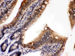
- IHC analysis of PDPK1 using anti-PDPK1 antibody (PB9735). PDPK1 was detected in paraffin-embedded section of Mouse Intestine Tissue. Heat mediated antigen retrieval was performed in citrate buffer (pH6, epitope retrieval solution) for 20 mins. The tissue section was blocked with 10% goat serum. The tissue section was then incubated with 1μg/ml rabbit anti-PDPK1 Antibody (PB9735) overnight at 4°C. Biotinylated goat anti-rabbit IgG was used as secondary antibody and incubated for 30 minutes at 37°C. The tissue section was developed using Strepavidin-Biotin-Complex (SABC)(Catalog # SA1022) with DAB as the chromogen.
- Additional image