Explore
 Validate
 Learn
Learn Western blot
 Immunocytochemistry
Antibody data
- Antibody Data
- Antigen structure
- References [6]
- Comments [0]
- Validations
- Immunocytochemistry [4]
- Immunoprecipitation [1]
- Immunohistochemistry [1]
- Other assay [3]
Submit
Validation data
Reference
Comment
Report error
- Product number
- MA1-010 - Provider product page

- Provider
- Invitrogen Antibodies
- Product name
- TRAP1 Monoclonal Antibody (TRAP1-6)
- Antibody type
- Monoclonal
- Antigen
- Purifed from natural sources
- Description
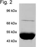
- MA1-010 detects tumor necrosis factor receptor-associated protein (TRAP1) from human tissues. MA1-010 has been successfully used in Western blot, immunofluorescence and immunoprecipitation procedures. By Western blot, this antibody detects an ~75 kDa protein representing TRAP1. Immunofluorescence staining of TRAP1 in PC-3-M cells with MA1-010 produces a pattern consistent with mitochondrial staining. Immunoprecipitation of TRAP1 using MA1-010 fails to co-precipitate p23, Hop, or CyP40 suggesting TRAP1s inability to associate with these co-chaperones. The MA1-010 immunogen is purified, recombinant, human TRAP1.
- Reactivity
- Human
- Host
- Mouse
- Isotype
- IgG
- Antibody clone number
- TRAP1-6
- Vial size
- 100 μg
- Concentration
- 1 mg/mL
- Storage
- -20°C, Avoid Freeze/Thaw Cycles
Submitted references Mitochondria are devoid of poly(ADP-ribose)polymerase-1, but harbor its product oligo(ADP-ribose).
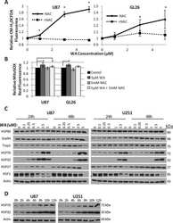
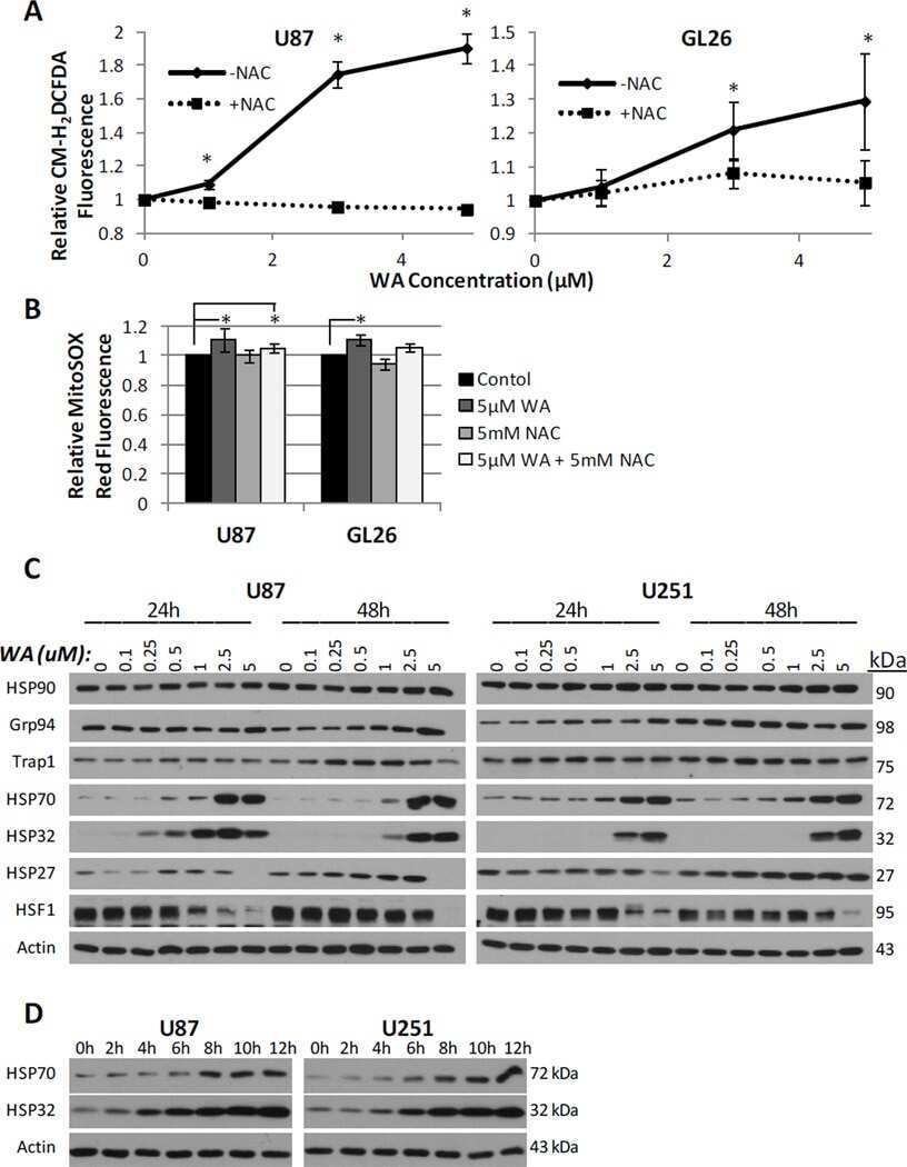
Cytotoxicity of withaferin A in glioblastomas involves induction of an oxidative stress-mediated heat shock response while altering Akt/mTOR and MAPK signaling pathways.
A novel C-terminal HSP90 inhibitor KU135 induces apoptosis and cell cycle arrest in melanoma cells.
Involvement of tumor necrosis factor receptor-associated protein 1 (TRAP1) in apoptosis induced by beta-hydroxyisovalerylshikonin.
Hsp90 chaperone activity requires the full-length protein and interaction among its multiple domains.
The hsp90-related protein TRAP1 is a mitochondrial protein with distinct functional properties.
Köritzer J, Blenn C, Bürkle A, Beneke S
Journal of cellular biochemistry 2021 May;122(5):507-523
Journal of cellular biochemistry 2021 May;122(5):507-523
Cytotoxicity of withaferin A in glioblastomas involves induction of an oxidative stress-mediated heat shock response while altering Akt/mTOR and MAPK signaling pathways.
Grogan PT, Sleder KD, Samadi AK, Zhang H, Timmermann BN, Cohen MS
Investigational new drugs 2013 Jun;31(3):545-57
Investigational new drugs 2013 Jun;31(3):545-57
A novel C-terminal HSP90 inhibitor KU135 induces apoptosis and cell cycle arrest in melanoma cells.
Samadi AK, Zhang X, Mukerji R, Donnelly AC, Blagg BS, Cohen MS
Cancer letters 2011 Dec 22;312(2):158-67
Cancer letters 2011 Dec 22;312(2):158-67
Involvement of tumor necrosis factor receptor-associated protein 1 (TRAP1) in apoptosis induced by beta-hydroxyisovalerylshikonin.
Masuda Y, Shima G, Aiuchi T, Horie M, Hori K, Nakajo S, Kajimoto S, Shibayama-Imazu T, Nakaya K
The Journal of biological chemistry 2004 Oct 8;279(41):42503-15
The Journal of biological chemistry 2004 Oct 8;279(41):42503-15
Hsp90 chaperone activity requires the full-length protein and interaction among its multiple domains.
Johnson BD, Chadli A, Felts SJ, Bouhouche I, Catelli MG, Toft DO
The Journal of biological chemistry 2000 Oct 20;275(42):32499-507
The Journal of biological chemistry 2000 Oct 20;275(42):32499-507
The hsp90-related protein TRAP1 is a mitochondrial protein with distinct functional properties.
Felts SJ, Owen BA, Nguyen P, Trepel J, Donner DB, Toft DO
The Journal of biological chemistry 2000 Feb 4;275(5):3305-12
The Journal of biological chemistry 2000 Feb 4;275(5):3305-12
No comments: Submit comment
Supportive validation
- Submitted by
- Invitrogen Antibodies (provider)
- Main image
- Experimental details
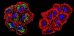
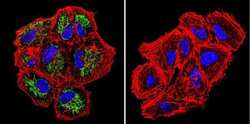
- Immunofluorescent analysis of TRAP1 in NCI-H460 Cells. Cells were grown on chamber slides and fixed with formaldehyde prior to staining. Cells were probed without (control) or with a TRAP1 monoclonal antibody (Product # MA1-010) at a dilution of 1:200 overnight at 4 C, washed with PBS and incubated with a DyLight-488 conjugated secondary antibody (Product # 35503). TRAP1 staining (green), F-Actin staining with Phalloidin (red) and nuclei with DAPI (blue) is shown. Images were taken at 60X magnification.

- Submitted by
- Invitrogen Antibodies (provider)
- Main image
- Experimental details
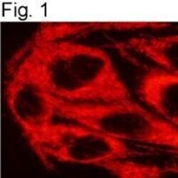
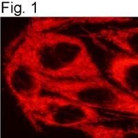
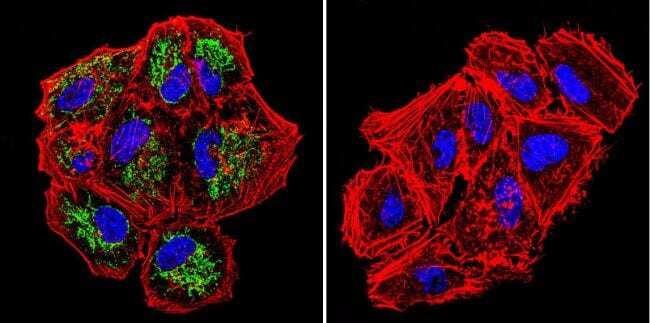
- Immunofluorescent analysis of TRAP1 in PC-3-M cells using a TRAP1 monoclonal antibody (Product # MA1-010).
- Submitted by
- Invitrogen Antibodies (provider)
- Main image
- Experimental details
- Immunofluorescent analysis of TRAP1 in NCI-H460 Cells. Cells were grown on chamber slides and fixed with formaldehyde prior to staining. Cells were probed without (control) or with a TRAP1 monoclonal antibody (Product # MA1-010) at a dilution of 1:200 overnight at 4 C, washed with PBS and incubated with a DyLight-488 conjugated secondary antibody (Product # 35503). TRAP1 staining (green), F-Actin staining with Phalloidin (red) and nuclei with DAPI (blue) is shown. Images were taken at 60X magnification.
- Submitted by
- Invitrogen Antibodies (provider)
- Main image

- Experimental details
- Immunofluorescent analysis of TRAP1 in PC-3-M cells using a TRAP1 monoclonal antibody (Product # MA1-010).

Supportive validation
- Submitted by
- Invitrogen Antibodies (provider)
- Main image

- Experimental details
- Immunoprecipitation of TRAP1 using Product # MA1-010 visualized by Coomassie Blue staining.

Supportive validation
- Submitted by
- Invitrogen Antibodies (provider)
- Main image
- Experimental details
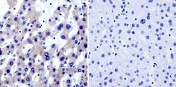
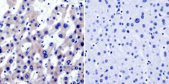
- Immunohistochemistry was performed on normal biopsies of deparaffinized Human liver tissue. To expose target proteins, heat induced antigen retrieval was performed using 10mM sodium citrate (pH6.0) buffer, microwaved for 8-15 minutes. Following antigen retrieval tissues were blocked in 3% BSA-PBS for 30 minutes at room temperature and probed with a TRAP1 monoclonal antibody (Product # MA1-010) at a dilution of 1:20 or without primary antibody (negative control) overnight at 4°C in a humidified chamber. Tissues were washed with PBST and endogenous peroxidase activity was quenched with a peroxidase suppressor. Detection was performed using a biotin-conjugated secondary antibody and SA-HRP, followed by colorimetric detection using DAB. Tissues were counterstained with hematoxylin and prepped for mounting.
Supportive validation
- Submitted by
- Invitrogen Antibodies (provider)
- Main image
- Experimental details
- NULL
- Submitted by
- Invitrogen Antibodies (provider)
- Main image
- Experimental details
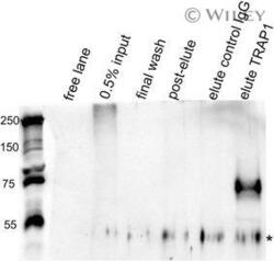
- 6 Figure TRAP1 interacts with oADP-ribose in mitochondria. Mitochondria isolated from 10 8 HeLaS3 cells were immunoprecipitated using TRAP1 (75 kDa) antibody. 0.5% input, 5% of final wash, 25% post-elute, and 40% of eluted material of IP either with an unrelated IgG antibody (elute control IgG) or with TRAP1 antibody (elute) were probed for oADPR presence using LP96-10. Whereas the high molecular weight signal is visible in the input fraction, this is no longer detected in the TRAP1-IP elute, but the specific signal at 75 kDa (arrow). * denotes signals from mouse IgG used for IP. IP, immunoprecipitation; PARP, poly(ADP-ribose)polymerase

- Submitted by
- Invitrogen Antibodies (provider)
- Main image

- Experimental details
- Immunoprecipitation of TRAP1 using Product # MA1-010 visualized by Coomassie Blue staining.