 Explore
 Validate
 Learn
Learn Western blot
Antibody data
- Antibody Data
- Antigen structure
- References [0]
- Comments [0]
- Validations
- Western blot [3]
- Immunocytochemistry [2]
Submit
Validation data
Reference
Comment
Report error
- Product number
- PA5-21826 - Provider product page

- Provider
- Invitrogen Antibodies
- Product name
- beta Tubulin Polyclonal Antibody
- Antibody type
- Polyclonal
- Antigen
- Recombinant protein fragment
- Description
- Recommended positive controls: 293T, A431, H1299, HeLaS3, HepG2, Molt-4, Raji, NIH-3T3. Predicted reactivity: Mouse (92%), Cat (81%), Pig (90%), Chicken (86%), Bovine (93%). Store product as a concentrated solution. Centrifuge briefly prior to opening the vial.
- Reactivity
- Human, Mouse, Rat
- Host
- Rabbit
- Isotype
- IgG
- Vial size
- 100 µL
- Concentration
- 1 mg/mL
- Storage
- Store at 4°C short term. For long term storage, store at -20°C, avoiding freeze/thaw cycles.
No comments: Submit comment
Supportive validation
- Submitted by
- Invitrogen Antibodies (provider)
- Main image
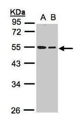
- Experimental details
- Western Blot using beta Tubulin Polyclonal Antibody (Product # PA5-21826). Sample (30 µg whole cell lysate). A: A431. B: Hep G2. 10% SDS PAGE. Beta Tubulin Polyclonal Antibody (Product # PA5-21826) diluted at 1:1,000.
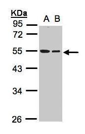
- Submitted by
- Invitrogen Antibodies (provider)
- Main image
- Experimental details
- Western Blot using beta Tubulin Polyclonal Antibody (Product # PA5-21826). Sample (30 µg of whole cell lysate). Lane A: NIH-3T3. 10% SDS PAGE. beta Tubulin Polyclonal Antibody (Product # PA5-21826) diluted at 1:1,000.

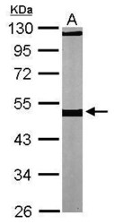
- Submitted by
- Invitrogen Antibodies (provider)
- Main image

- Experimental details
- Western blot was performed using Anti-beta Tubulin Rabbit Polyclonal Antibody (Product # PA5-21826) and a 52kDa band corresponding to beta Tubulin was observed across cell lines tested. Whole cell extracts (30 µg lysate) of Hep G2 (Lane 1), NIH-3T3 (Lane 2) and PC-12 (Lane 3) were electrophoresed using Novex® NuPAGE® 4-12 % Bis-Tris gel (Product # NP0322BOX). Resolved proteins were then transferred onto a nitrocellulose membrane (Product # IB23001) by iBlot® 2 Dry Blotting System (Product # IB21001). The blot was probed with the primary antibody (1:2000 dilution) and detected by chemiluminescence with Goat anti-Rabbit IgG (H+L), Superclonal™ Recombinant Secondary Antibody, HRP (Product # A27036, 1:4000 dilution) using the iBright FL 1000 (Product # A32752). Chemiluminescent detection was performed using Novex® ECL Chemiluminescent Substrate Reagent Kit (Product # WP20005).

Supportive validation
- Submitted by
- Invitrogen Antibodies (provider)
- Main image

- Experimental details
- Immunocytochemistry-Immunofluorescence analysis of beta Tubulin was performed in HeLa cells fixed in 4% paraformaldehyde at RT for 15 min. Green: beta Tubulin Polyclonal Antibody (Product # PA5 21826) diluted at 1:200. Blue: Hoechst 33342 staining. Scale bar = 10 µm.

- Submitted by
- Invitrogen Antibodies (provider)
- Main image

- Experimental details
- Immunofluorescence analysis of beta Tubulin was performed using 70% confluent log phase HeLa cells. The cells were fixed with 4% paraformaldehyde for 10 minutes, permeabilized with 0.1% Triton™ X-100 for 15 minutes, and blocked with 1% BSA for 1 hour at room temperature. The cells were labeled with beta Tubulin Polyclonal Antibody (Product # PA5-21826) at 1:100 dilution in 0.1% BSA, incubated at 4 degree Celsius overnight and then labeled with Goat anti-Rabbit IgG (H+L), Superclonal™ Recombinant Secondary Antibody, Alexa Fluor 488 (Product # A27034) at a dilution of 1:2000 for 45 minutes at room temperature (Panel a: green). Nuclei (Panel b: blue) were stained with SlowFade® Gold Antifade Mountant with DAPI (Product # S36938). F-actin (Panel c: red) was stained with Rhodamine Phalloidin (Product # R415, 1:300). Panel d represents the merged image showing microtubular localization. Panel e represents control cells with no primary antibody to assess background. The images were captured at 60X magnification.
