 Explore
 Validate
 Learn
Learn Western blot
 Immunocytochemistry
Antibody data
- Antibody Data
- Antigen structure
- References [0]
- Comments [0]
- Validations
- Immunocytochemistry [3]
- Immunohistochemistry [4]
- Flow cytometry [2]
Submit
Validation data
Reference
Comment
Report error
- Product number
- MA5-34809 - Provider product page

- Provider
- Invitrogen Antibodies
- Product name
- RACK1 Recombinant Rabbit Monoclonal Antibody (JG40-26)
- Antibody type
- Monoclonal
- Antigen
- Recombinant full-length protein
- Description
- Positive Control: SH-SY-5Y, rat small intestine tissue lysate, rat cerebellum tissue, rat liver tissue, human colon tissue, mouse skin tissue.
- Reactivity
- Human, Mouse, Rat
- Host
- Rabbit
- Isotype
- IgG
- Antibody clone number
- JG40-26
- Vial size
- 100 μL
- Concentration
- 1 mg/mL
- Storage
- -20°C, Avoid Freeze/Thaw Cycles, store in dark
No comments: Submit comment
Supportive validation
- Submitted by
- Invitrogen Antibodies (provider)
- Main image
- Experimental details
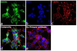
- Immunofluorescence analysis of RACK1 was performed using 70% confluent log phase HEK-293 cells. The cells were fixed with 4% paraformaldehyde for 10 minutes, permeabilized with 0.1% Triton™ X-100 for 10 minutes, and blocked with 2% BSA for 1 hour at room temperature. The cells were labeled with RACK1 Recombinant Rabbit Monoclonal Antibody (JG40-26) (Product # MA5-34809) at 1:100 in 0.1% BSA, incubated at 4 degree celsius overnight and then labeled with Goat anti-Rabbit IgG (Heavy Chain) Superclonal™ Recombinant Secondary Antibody, Alexa Fluor® 488 conjugate (Product # A27034), (1:2000), for 45 minutes at room temperature (Panel a: Green). Nuclei (Panel b:Blue) were stained with Hoechst 33342 (Product # H1399). F-actin (Panel c: Red) was stained with Alexa Fluor™ Plus 647 Phalloidin (Product # A30107, 1:2000 dilution). Panel d represents the merged image showing nucleus, cytoplasm localization. Panel e represents control cells with no primary antibody to assess background. The images were captured at 40X magnification. The images were captured at 40X magnification in CellInsight CX7 LZR High-Content Screening (HCS) Platform (Product # CX7A1110LZR) and externally deconvoluted (D.Sage et al./Methods 115 (2017) 28–41.
- Submitted by
- Invitrogen Antibodies (provider)
- Main image

- Experimental details
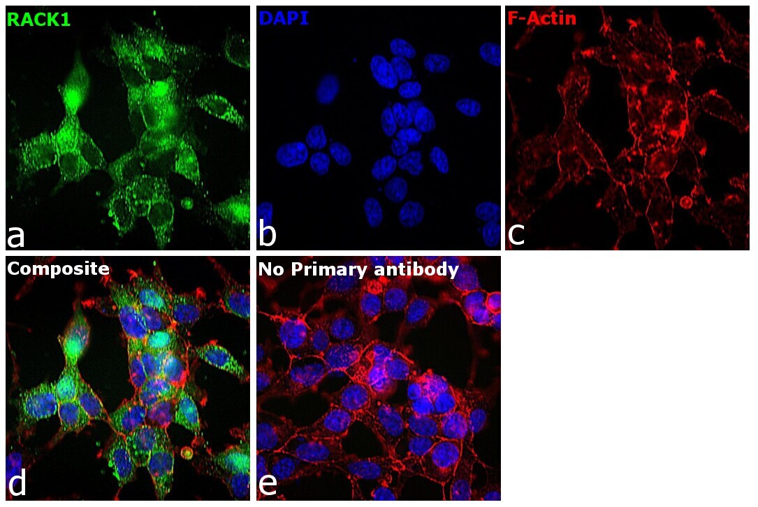
- Immunofluorescence analysis of RACK1 was performed using 70% confluent log phase HeLa cells. The cells were fixed with 4% paraformaldehyde for 10 minutes, permeabilized with 0.1% Triton™ X-100 for 10 minutes, and blocked with 2% BSA for 1 hour at room temperature. The cells were labeled with RACK1 Recombinant Rabbit Monoclonal Antibody (JG40-26) (Product # MA5-34809) at 1:100 in 0.1% BSA, incubated at 4 degree celsius overnight and then labeled with Goat anti-Rabbit IgG (H+L) Superclonal™ Recombinant Secondary Antibody, Alexa Fluor® 488 conjugate (Product # A27034), (1:2000), for 45 minutes at room temperature (Panel a: Green). Nuclei (Panel b:Blue) were stained with Hoechst 33342 (Product # H1399). F-actin (Panel c: Red) was stained with Alexa Fluor™ Plus 647 Phalloidin (Product # A30107, 1:2000 dilution). Panel d represents the merged image showing nucleus, cytoplasm localization. Panel e represents control cells with no primary antibody to assess background. The images were captured at 40X magnification. The images were captured at 40X magnification in CellInsight CX7 LZR High-Content Screening (HCS) Platform (Product # CX7A1110LZR) and externally deconvoluted (D.Sage et al./Methods 115 (2017) 28–41.
- Submitted by
- Invitrogen Antibodies (provider)
- Main image
- Experimental details
- Immunofluorescence analysis of RACK1 was performed using 70% confluent log phase HeLa cells. The cells were fixed with 4% paraformaldehyde for 10 minutes, permeabilized with 0.1% Triton™ X-100 for 10 minutes, and blocked with 2% BSA for 1 hour at room temperature. The cells were labeled with RACK1 Recombinant Rabbit Monoclonal Antibody (JG40-26) (Product # MA5-34809) at 1:100 in 0.1% BSA, incubated at 4 degree celsius overnight and then labeled with Goat anti-Rabbit IgG (Heavy Chain) Superclonal™ Recombinant Secondary Antibody, Alexa Fluor® 488 conjugate (Product # A27034), (1:2000), for 45 minutes at room temperature (Panel a: Green). Nuclei (Panel b:Blue) were stained with Hoechst 33342 (Product # H1399). F-actin (Panel c: Red) was stained with Alexa Fluor™ Plus 647 Phalloidin (Product # A30107, 1:2000 dilution). Panel d represents the merged image showing nucleus, cytoplasm localization. Panel e represents control cells with no primary antibody to assess background. The images were captured at 40X magnification. The images were captured at 40X magnification in CellInsight CX7 LZR High-Content Screening (HCS) Platform (Product # CX7A1110LZR) and externally deconvoluted (D.Sage et al./Methods 115 (2017) 28–41.

Supportive validation
- Submitted by
- Invitrogen Antibodies (provider)
- Main image
- Experimental details
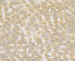
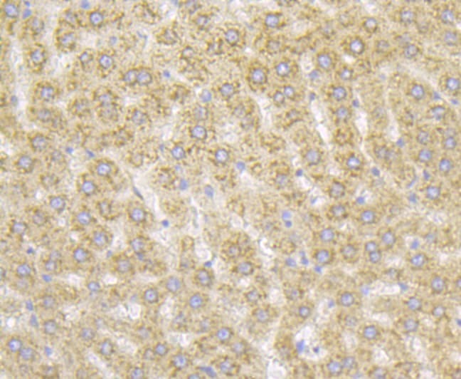
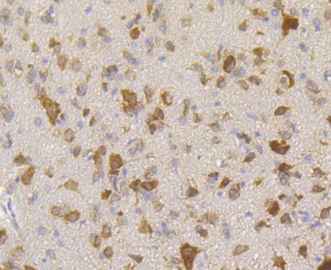
- Immunohistochemistry analysis of RACK1 in paraffin-embedded rat liver tissue. Samples were incubated with RACK1 monoclonal antibody (Product # MA5-34809), and followed by hematoxylin.
- Submitted by
- Invitrogen Antibodies (provider)
- Main image
- Experimental details
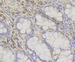
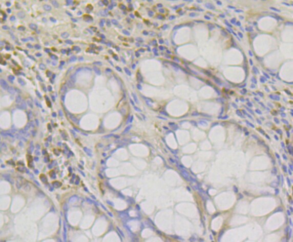
- Immunohistochemistry analysis of RACK1 in paraffin-embedded human colon tissue. Samples were incubated with RACK1 monoclonal antibody (Product # MA5-34809), and followed by hematoxylin.
- Submitted by
- Invitrogen Antibodies (provider)
- Main image
- Experimental details
- Immunohistochemistry analysis of RACK1 in paraffin-embedded mouse skin tissue. Samples were incubated with RACK1 monoclonal antibody (Product # MA5-34809), and followed by hematoxylin.

- Submitted by
- Invitrogen Antibodies (provider)
- Main image
- Experimental details
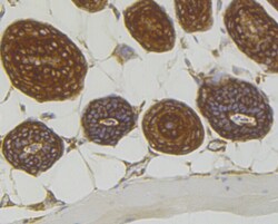
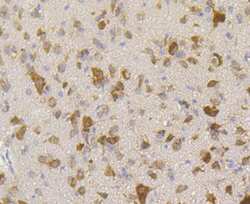
- Immunohistochemistry analysis of RACK1 in paraffin-embedded rat cerebellum tissue. Samples were incubated with RACK1 monoclonal antibody (Product # MA5-34809), and followed by hematoxylin.
Supportive validation
- Submitted by
- Invitrogen Antibodies (provider)
- Main image
- Experimental details
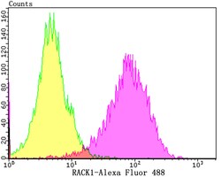
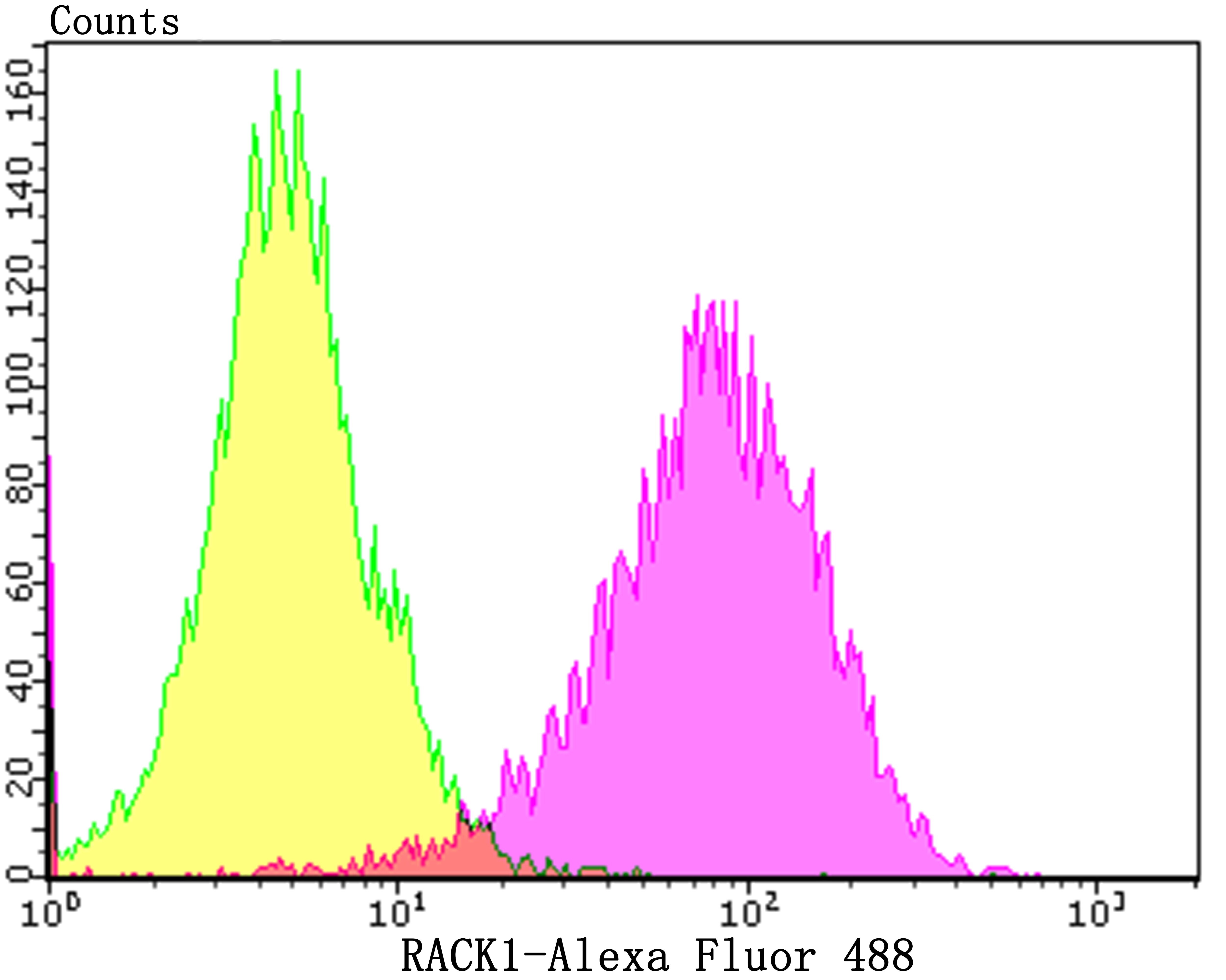
- Flow cytometry of RACK1 in SH-SY-5Y cells (purple) compared with an unlabelled control (cells without incubation with primary antibody; yellow). Samples were incubated with RACK1 monoclonal antibody (Product # MA5-34809) at a dilution of 1:100, followed by Alexa Fluor 488-conjugated goat anti-rabbit IgG.
- Submitted by
- Invitrogen Antibodies (provider)
- Main image

- Experimental details
- Flow cytometry of RACK1 in SH-SY-5Y cells (purple) compared with an unlabelled control (cells without incubation with primary antibody; yellow). Samples were incubated with RACK1 monoclonal antibody (Product # MA5-34809) at a dilution of 1:100, followed by Alexa Fluor 488-conjugated goat anti-rabbit IgG.
