 Explore
 Validate
 Learn
Learn Western blot
Antibody data
- Antibody Data
- Antigen structure
- References [2]
- Comments [0]
- Validations
- Western blot [1]
- Other assay [2]
Submit
Validation data
Reference
Comment
Report error
- Product number
- PA5-37474 - Provider product page

- Provider
- Invitrogen Antibodies
- Product name
- Phospho-Annexin A2 (Ser26) Polyclonal Antibody
- Antibody type
- Polyclonal
- Antigen
- Synthetic peptide
- Description
- A suggested positive control for Western blot is HepG2 or JK cells. The homology between synthetic peptide sequence and Mouse/Rat is 100%.
- Reactivity
- Human
- Host
- Rabbit
- Isotype
- IgG
- Vial size
- 100 µL
- Concentration
- 1 mg/mL
- Storage
- -20°C
Submitted references Dephosphorylation of annexin A2 by protein phosphatase 1 regulates endothelial cell barrier.
Clinical Significance of Annexin A2 Expression in Breast Cancer Patients.
Király N, Thalwieser Z, Fonódi M, Csortos C, Boratkó A
IUBMB life 2021 Oct;73(10):1257-1268
IUBMB life 2021 Oct;73(10):1257-1268
Clinical Significance of Annexin A2 Expression in Breast Cancer Patients.
Gibbs LD, Mansheim K, Maji S, Nandy R, Lewis CM, Vishwanatha JK, Chaudhary P
Cancers 2020 Dec 22;13(1)
Cancers 2020 Dec 22;13(1)
No comments: Submit comment
Supportive validation
- Submitted by
- Invitrogen Antibodies (provider)
- Main image
- Experimental details
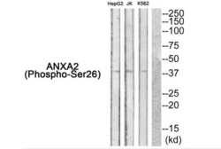
- Western blot analysis of extracts from HepG2 cells JK cells and K562 cells using ANXA2 (pSer26) polyclonal antibody (Product # PA5-37474) (left) or the same antibody preincubated with a blocking peptide (right).

Supportive validation
- Submitted by
- Invitrogen Antibodies (provider)
- Main image

- Experimental details
- 2 FIGURE PKC phosphorylates ANXA2 on Ser25 side chain. (a) Purified recombinant GST, GST-ANXA2, and GST-TIMAP proteins were incubated with or without active PKCalpha for 30 min. Amount of loaded proteins were verified by SDS-PAGE followed by Coomassie staining. Phosphorylation of proteins was analyzed by western blot using anti-phosphoSer PKC substrate and phospho-Ser25 annexin A2-specific antibodies. (b) Pull down of endothelial cell lysate was made using GST, GST-tagged TIMAP wild type, S331D or S331D recombinant full-length TIMAP proteins as baits. Samples were tested for ANXA2 in western blot experiment. (c) Total cell lysates of control, nonsilencing RNA and PKC-specific siRNA transfected HPAEC treated with PMA (1 muM for 30 min) or PMA (1 muM for 30 min) after a pretreatment with Go6983 (1 muM for 30 min) were analyzed by western blot using PKCalpha, actin, ANXA2, and phospho-Ser25 ANXA2-specific antibodies. (d) Western blot analysis of control, PMA (1 muM, 30 min), PMA (1 muM, 30 min) with Go6983 pretreatment (1 muM, 30 min), okadaic acid (5 nM, 30 min), tautomycetin (1 muM, 30 min), nonsiRNA, or TIMAP-specific siRNA-treated BPAEC cells are shown. Phospho-Ser25 ANXA2 level was normalized to ANXA2 protein level. Densitometric analysis of western blots are shown as mean +- SD ( n = 3). Statistical analysis was done with ANOVA and marked by asterisks; *** p < .001

- Submitted by
- Invitrogen Antibodies (provider)
- Main image

- Experimental details
- Figure 5 Phosphorylation of AnxA2 in breast cancer cells. ( A ) The expression of pAnxA2-S25, pAnxA2-Y23, and AnxA2 were analyzed in non-tumorigenic mammary epithelial cells and TNBC cells by immunoblot analysis. The membrane was stripped and reprobed with anti-beta-actin antibody for loading control. ( B ) Bar chart showing the densitometric analysis of pAnxA2-S25, pAnxA2-Y23 and AnxA2 bands of the immunoblot of panel A. Intensity of each bands were normalized by loading control. Each bar represents the mean +- SE of three independent experiments. ( C ) The HCC1395 cells were transfected with a plasmid vector expressing GFP alone, AnxA2-WT-GFP, and AnxA2-Y23F-GFP. The expression of pAnxA2-Y23, AnxA2 and GFP were analyzed by immunoblot analysis. The membrane was stripped and reprobed with anti-beta-actin antibody for loading control. ( D ) The HCC1395 transfected cells (6 x 10 5 ) were plated in 100mm Petri dish for overnight and then switched to serum free medium. After 24 h, the AnxA2-GFP complex was immunoprecipitated from the medium using anti-GFP antibody (Catalog no. DSHB-GFP-12A6; DSHB) and expression of AnxA2 secretion was analyzed by anti-AnxA2 antibody using immunoblot analysis. Uncropped Western Blots of ( A , C , D ) are available in Figure S3 .
