Explore
 Validate
 Learn
Learn Western blot
 Other assay
Antibody data
- Antibody Data
- Antigen structure
- References [2]
- Comments [0]
- Validations
- Other assay [2]
Submit
Validation data
Reference
Comment
Report error
- Product number
- PA1-9006 - Provider product page

- Provider
- Invitrogen Antibodies
- Product name
- Annexin A2 Polyclonal Antibody
- Antibody type
- Polyclonal
- Antigen
- Synthetic peptide
- Description
- This antibody is predicted to react with bovine, porcine and rat based on sequence homology. This antibody is tested in Peptide ELISA: antibody detection limit dilution 1,000.
- Reactivity
- Human, Mouse
- Host
- Goat
- Isotype
- IgG
- Vial size
- 100 μg
- Concentration
- 0.5 mg/mL
- Storage
- -20°C, Avoid Freeze/Thaw Cycles
Submitted references The Tick Protein Sialostatin L2 Binds to Annexin A2 and Inhibits NLRC4-Mediated Inflammasome Activation.
Analysis of the effect of a novel therapeutic for type 2 diabetes on the proteome of a muscle cell line.
Wang X, Shaw DK, Sakhon OS, Snyder GA, Sundberg EJ, Santambrogio L, Sutterwala FS, Dumler JS, Shirey KA, Perkins DJ, Richard K, Chagas AC, Calvo E, Kopecký J, Kotsyfakis M, Pedra JHF
Infection and immunity 2016 Jun;84(6):1796-1805
Infection and immunity 2016 Jun;84(6):1796-1805
Analysis of the effect of a novel therapeutic for type 2 diabetes on the proteome of a muscle cell line.
Young PA, Leonard S, Martin DS, Findlay JB
Proteomics 2016 Jan;16(1):70-9
Proteomics 2016 Jan;16(1):70-9
No comments: Submit comment
Supportive validation
- Submitted by
- Invitrogen Antibodies (provider)
- Main image
- Experimental details
- NULL
- Submitted by
- Invitrogen Antibodies (provider)
- Main image

- Experimental details
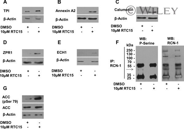
- Secondary validation of some of the changes observed in response to RTC-15 treatment. Western blotting analysis of (A) TPI, (B) annexin A2, (C) calumenin, (D) ZPR1, and (E) ECH1 expression. All experiments were conducted in triplicate. beta-Actin was used as a loading control where appropriate. As no phosphospecific antibodies were available for RCN-1, the protein was first immunoprecipitated and subsequently subjected to Western blotting analysis. Serine phosphorylation was assessed using anti-phosphoserine antibody and total protein levels were observed using anti-RCN-1 antibody (F). Change in ACC phosphorylation in response to RTC-15 (G).