Explore
 Validate
 Learn
Learn Western blot
Antibody data
- Antibody Data
- Antigen structure
- References [1]
- Comments [0]
- Validations
- Western blot [3]
- Immunohistochemistry [1]
Submit
Validation data
Reference
Comment
Report error
- Product number
- AF3928 - Provider product page

- Provider
- R&D Systems
- Product name
- Human/Mouse/Rat Annexin A2 Antibody
- Antibody type
- Polyclonal
- Description
- Antigen Affinity-purified. Detects human, mouse, and rat Annexin A2 in Western blots. In Western blots, no cross-reactivity with recombinant human Annexin A1, A3, A4, A5, A6, A7, A8, A9, A10, A11, or A13 is observed.
- Reactivity
- Human, Mouse, Rat
- Host
- Goat
- Conjugate
- Unconjugated
- Antigen sequence
P07355- Isotype
- IgG
- Vial size
- 100 ug
- Concentration
- LYOPH
- Storage
- Use a manual defrost freezer and avoid repeated freeze-thaw cycles. 12 months from date of receipt, -20 to -70 °C as supplied. 1 month, 2 to 8 °C under sterile conditions after reconstitution. 6 months, -20 to -70 °C under sterile conditions after reconstitution.
Submitted references The hemopexin domain of MMP3 is responsible for mammary epithelial invasion and morphogenesis through extracellular interaction with HSP90β.
Correia AL, Mori H, Chen EI, Schmitt FC, Bissell MJ
Genes & development 2013 Apr 1;27(7):805-17
Genes & development 2013 Apr 1;27(7):805-17
No comments: Submit comment
Supportive validation
- Submitted by
- R&D Systems (provider)
- Main image

- Experimental details
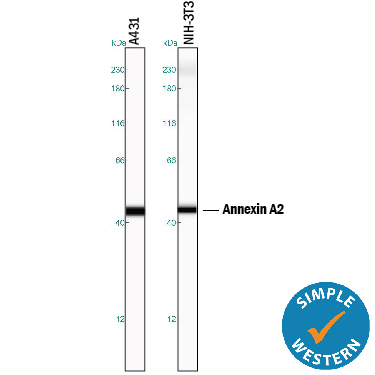
- Detection of Human and Mouse Annexin A2 by Simple WesternTM. Simple Western lane view shows lysates of A431 human epithelial carcinoma cell line and NIH-3T3 mouse embryonic fibroblast cell line, loaded at 0.2 mg/mL. A specific band was detected for Annexin A2 at approximately 45 kDa (as indicated) using 50 µg/mL of Goat Anti-Human/Mouse/Rat Annexin A2 Antigen Affinity-purified Polyclonal Antibody (Catalog # AF3928) followed by 1:50 dilution of HRP-conjugated Anti-Goat IgG Secondary Antibody (Catalog # HAF109). This experiment was conducted under reducing conditions and using the 12-230 kDa separation system. Non-specific interaction with the 230 kDa Simple Western standard may be seen with this antibody.
- Submitted by
- R&D Systems (provider)
- Main image
- Experimental details
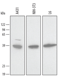
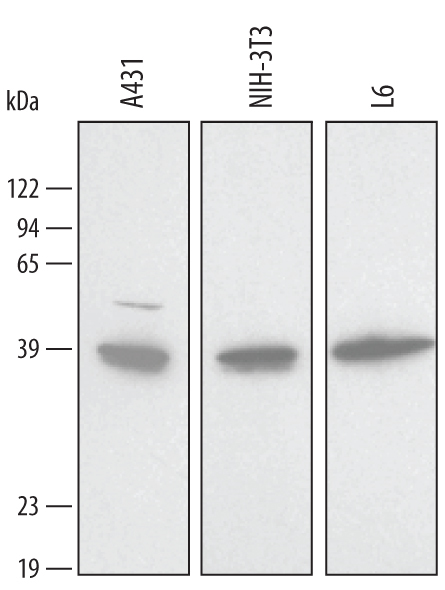
- Detection of Human/Mouse/Rat Annexin A2 by Western Blot. Western blot shows lysates of A431 human epithelial carcinoma cell line, NIH-3T3 mouse embryonic fibroblast cell line, and L6 rat myoblast cell line. PVDF membrane was probed with 1 µg/mL of Goat Anti-Human/Mouse/Rat Annexin A2 Antigen Affinity-purified Polyclonal Antibody (Catalog # AF3928) followed by HRP-conjugated Anti-Goat IgG Secondary Antibody (Catalog # HAF017). A specific band was detected for Annexin A2 at approximately 39 kDa (as indicated). This experiment was conducted using Immunoblot Buffer Group 2.
- Submitted by
- R&D Systems (provider)
- Main image
- Experimental details
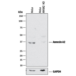
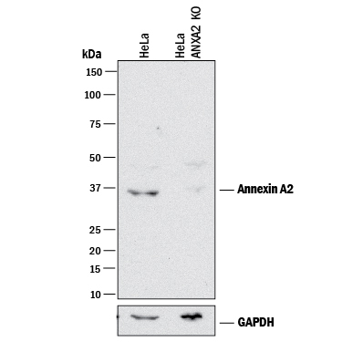
- Western Blot Shows Human Annexin A2 Specificity by Using Knockout Cell Line. Western blot shows lysates of HeLa human cervical epithelial carcinoma parental cell line and Annexin A2 knockout HeLa cell line (KO). PVDF membrane was probed with 1 µg/mL of Goat Anti-Human/Mouse/Rat Annexin A2 Antigen Affinity-purified Polyclonal Antibody (Catalog # AF3928) followed by HRP-conjugated Anti-Goat IgG Secondary Antibody (Catalog # HAF017). A specific band was detected for Annexin A2 at approximately 37 kDa (as indicated) in the parental HeLa cell line, but is not detectable in knockout HeLa cell line. GAPDH (Catalog # AF5718) is shown as a loading control. This experiment was conducted under reducing conditions and using Immunoblot Buffer Group 1.
Supportive validation
- Submitted by
- R&D Systems (provider)
- Main image
- Experimental details
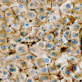
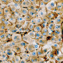
- Annexin A2 in Human Liver. Annexin A2 was detected in immersion fixed paraffin-embedded sections of normal human liver using Goat Anti-Human/Mouse/Rat Annexin A2 Antigen Affinity-purified Polyclonal Antibody (Catalog # AF3928) at 3 µg/mL overnight at 4 °C. Before incubation with the primary antibody, tissue was subjected to heat-induced epitope retrieval using Antigen Retrieval Reagent-Basic (Catalog # CTS013). Tissue was stained using the Anti-Goat HRP-DAB Cell & Tissue Staining Kit (brown; Catalog # CTS008) and counterstained with hematoxylin (blue). Specific staining was localized to plasma membranes of hepatocytes. View our protocol for Chromogenic IHC Staining of Paraffin-embedded Tissue Sections.