Explore
 Validate
 Learn
Learn Western blot
Antibody data
- Antibody Data
- Antigen structure
- References [0]
- Comments [0]
- Validations
- Western blot [1]
- Immunohistochemistry [1]
- Flow cytometry [1]
Submit
Validation data
Reference
Comment
Report error
- Product number
- TA325068 - Provider product page

- Provider
- OriGene
- Product name
- Rabbit polyclonal ANXA7 Antibody (Center)
- Antibody type
- Polyclonal
- Description
- Rabbit polyclonal ANXA7 Antibody (Center)
- Host
- Rabbit
- Conjugate
- Unconjugated
- Epitope
- ANXA7
- Isotype
- IgG
- Antibody clone number
- NULL
- Vial size
- 400 µl
- Concentration
- 0.5 mg/ml
No comments: Submit comment
Supportive validation
- Submitted by
- OriGene (provider)
- Main image
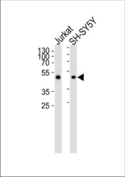
- Experimental details
- ANXA7 Antibody (Center) (Cat.# TA325068) western blot analysis in Jurkat and SH-SY5Y cell lysates (35ug/lane). This demonstrates that the ANXA7 antibody detected ANXA7 protein (arrow).
- Validation comment
- WB

Supportive validation
- Submitted by
- OriGene (provider)
- Main image

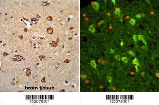
- Experimental details
- (LEFT)Formalin-fixed and paraffin-embedded human brain tissue reacted with ANXA7 Antibody (Center), which was peroxidase-conjugated to the secondary antibody, followed by DAB staining. (RIGHT)Immunofluorescence analysis of ANXA7 Antibody (Center) with paraffin-embedded human brain tissue . 0.05 mg/ml primary antibody was followed by FITC-conjugated goat anti-rabbit lgG (whole molecule). FITC emits green fluorescence. Red counterstaining is PI.
- Validation comment
- IHC
Supportive validation
- Submitted by
- OriGene (provider)
- Main image
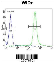
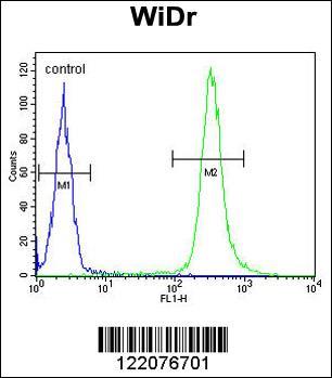
- Experimental details
- ANXA7 Antibody (Center) (Cat. #TA325068) flow cytometric analysis of WiDr cells (right histogram) compared to a negative control cell (left histogram).FITC-conjugated goat-anti-rabbit secondary antibodies were used for the analysis.
- Validation comment
- FC