Explore
 Validate
 Learn
Learn Western blot
Antibody data
- Antibody Data
- Antigen structure
- References [1]
- Comments [0]
- Validations
- Western blot [1]
- Immunocytochemistry [1]
- Flow cytometry [1]
Submit
Validation data
Reference
Comment
Report error
- Product number
- 702357 - Provider product page

- Provider
- Invitrogen Antibodies
- Product name
- Phospho-CaMKII (Thr305,Thr306) Recombinant Rabbit Monoclonal Antibody (9H2L7)
- Antibody type
- Monoclonal
- Antigen
- Synthetic peptide
- Description
- This antibody is predicted to react with Monkey, Mouse, Dog, Sheep and Bovine
- Antibody clone number
- 9H2L7
- Concentration
- 0.5 mg/mL
Submitted references Mitotic activity, modulation of DNA processing, and purinergic signalling in the adult rat auditory brainstem following sensory deafferentation.
Illing RB, Buschky H, Tadic A
The European journal of neuroscience 2019 Dec;50(12):3985-4003
The European journal of neuroscience 2019 Dec;50(12):3985-4003
No comments: Submit comment
Supportive validation
- Submitted by
- Invitrogen Antibodies (provider)
- Main image
- Experimental details
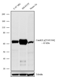
- Western blot analysis was performed on whole cell extracts (30 µg lysate) of U-87 MG (Lane 1), SH-SY5Y (Lane 2) and Neuro-2a (Lane 3). The blots were probed with Anti-CamKII p (T305/306) Recombinant Rabbit Monoclonal Antibody (Product # 702357, 1-2 µg/mL) and detected by chemiluminescence using Goat anti-Rabbit IgG (H+L) Superclonal™ Secondary Antibody, HRP conjugate (Product # A27036, 0.4 µg/mL, 1:2500 dilution). A 63 kDa band corresponding to CamKII p (T305/306) was observed across cell lines tested. Known quantity of protein samples were electrophoresed using Novex® NuPAGE® 4-12% Bis-Tris gel (Product # NP0321BOX), XCell SureLock™ Electrophoresis System (Product # EI0002) and Novex® Sharp Pre-Stained Protein Standard (Product # LC5800). Resolved proteins were then transferred onto a nitrocellulose membrane with iBlot® Dry Blotting System (Product # IB21001). The membrane was probed with the relevant primary and secondary Antibody following blocking with 5% skimmed milk. Chemiluminescent detection was performed using Pierce™ ECL Western blotting Substrate (Product # 32106).

Supportive validation
- Submitted by
- Invitrogen Antibodies (provider)
- Main image
- Experimental details
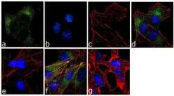
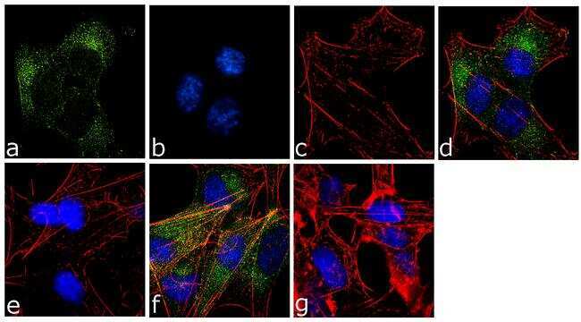
- For immunofluorescence analysis, SH-SY5Y cells were fixed and permeabilized for detection of endogenous CamKIIpT305/306 using Anti- CamKIIpT305/306 Recombinant Rabbit Monoclonal Antibody (Product # 702357, 2 µg/mL) and labeled with Goat anti-Rabbit IgG (H+L) Superclonal™ Secondary Antibody, Alexa Fluor® 488 conjugate (Product # A27034, 1:2000). Panel a) shows representative cells that were stained for detection and localization of CamKIIpT305/306 protein (green), Panel b) is stained for nuclei (blue) using SlowFade® Gold Antifade Mountant with DAPI (Product # S36938). Panel c) represents cytoskeletal F-actin staining using Alexa Fluor® 555 Rhodamine Phalloidin (Product # R415, 1:300). Panel d) is a composite image of Panels a, b and c clearly demonstrating cytoplasmic localization of CamKIIpT305/306. Panel e) shows loss of signal by competition with the CamKIIpT305/306 peptide, demonstrating antibody specificity. Panel f) demonstrates no competition with the non-phosphorylated peptide. Panel g) represents control cells with no primary antibody to assess background. The images were captured at 60X magnification.
Supportive validation
- Submitted by
- Invitrogen Antibodies (provider)
- Main image
- Experimental details
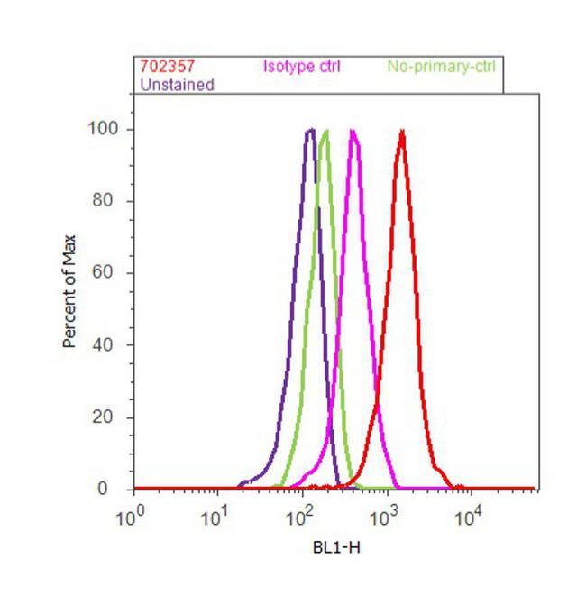
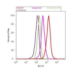
- Flow Cytometry analysis of endogenous CamKII (pT305/306) was performed on U-87 MG cells labeled with ABfinity™ Anti-CamKII (pT305/306) Recombinant Rabbit Monoclonal Antibody (Product# 702357, 5 ug/ 1M cells) or with rabbit isotype control at 0.5 ug/ml and detected with Goat anti-Rabbit IgG (H+L) Superclonal™ Secondary Antibody, (Alexa Fluor® 488 conjugate, Product# A27034, 0.4 ug/ml, 1:2500) as represented by the red and pink histograms respectively. The purple histogram represents unstained control cells and the green histogram represents no-primary-antibody control. A representative of 10,000 cells were acquired and analyzed for each sample using an Attune® Acoustic Focusing Cytometer (4468770).