Explore
 Validate
 Learn
Learn Western blot
 Immunocytochemistry
 Immunoprecipitation
Antibody data
- Antibody Data
- Antigen structure
- References [1]
- Comments [0]
- Validations
- Immunocytochemistry [3]
- Other assay [1]
Submit
Validation data
Reference
Comment
Report error
- Product number
- PA5-17453 - Provider product page

- Provider
- Invitrogen Antibodies
- Product name
- c-Cbl Polyclonal Antibody
- Antibody type
- Polyclonal
- Antigen
- Synthetic peptide
- Description
- It is not recommended to aliquot this antibody.
- Reactivity
- Human, Mouse, Rat
- Host
- Rabbit
- Isotype
- IgG
- Vial size
- 100 μL
- Concentration
- 14.5 μg/mL
- Storage
- -20°C
Submitted references c-Cbl Acts as an E3 Ligase Against DDA3 for Spindle Dynamics and Centriole Duplication during Mitosis.
Gwon D, Hong J, Jang CY
Molecules and cells 2019 Dec 31;42(12):840-849
Molecules and cells 2019 Dec 31;42(12):840-849
No comments: Submit comment
Supportive validation
- Submitted by
- Invitrogen Antibodies (provider)
- Main image
- Experimental details
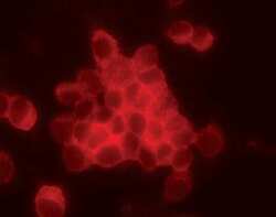
- Immunofluorescent analysis of c-Cbl in paraformaldehyde-fixed RAW cells, using a c-Cbl polyclonal antibody (Product # PA5-17453).
- Submitted by
- Invitrogen Antibodies (provider)
- Main image

- Experimental details
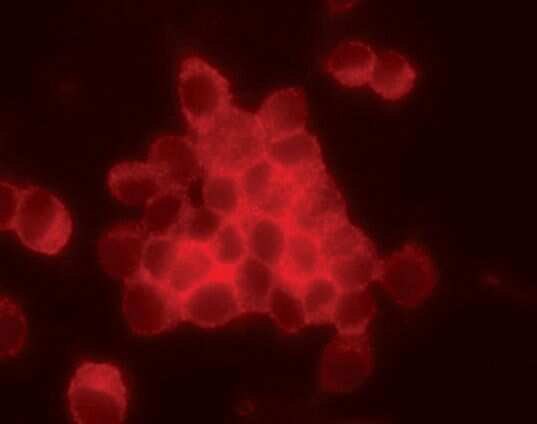
- Immunofluorescent analysis of c-Cbl in paraformaldehyde-fixed RAW cells, using a c-Cbl polyclonal antibody (Product # PA5-17453).

- Submitted by
- Invitrogen Antibodies (provider)
- Main image

- Experimental details
- Immunofluorescent analysis of c-Cbl in paraformaldehyde-fixed RAW cells, using a c-Cbl polyclonal antibody (Product # PA5-17453).

Supportive validation
- Submitted by
- Invitrogen Antibodies (provider)
- Main image
- Experimental details
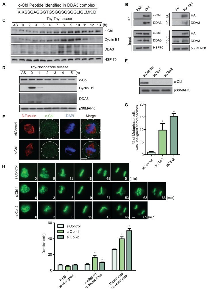
- Fig. 1 c-Cbl interacts with DDA3 and acts as a mitotic regulator (A) The DDA3 complex was purified from mitotic cells and analyzed by mass spectrometry. A peptide of c-Cbl was identified three times. (B) Twenty-eight hours after transfection of the EV or HA-c-Cbl plasmid, HeLa cells were harvested and subjected to immunoprecipitation and western blotting with the indicated antibodies. p38MAPK served as a loading control. EV, empty vector. (C and D) HeLa cells were synchronized by a double thymidine block (C) or thymidine-nocodazole block (D), placed into fresh media, and harvested at the indicated times. Cell lysates were analyzed by western blotting with the indicated antibodies. AS, unsynchronized cells. (E) HeLa cells were transfected with control (siControl) or c-Cbl-specific siRNAs (siCbl-A and siCbl-B). Seventy-two hours after siRNA transfection, the transfected cells were harvested and lysed to measure protein levels by western blotting with the indicated antibodies. (F and G) Seventy-two hours after siRNA transfection, HeLa cells were fixed with MeOH and stained with antibodies as indicated. Images are maximum projections from Z-stacks of representative cells stained for c-Cbl (green), beta-tubulin (red), and DNA (blue). The number of metaphase cells with unaligned chromosomes was quantified and plotted (G) (n = 300 metaphase cells from three independent experiments). (H) Seventy-two hours after siRNA transfection, HeLa cells expressing GFP-Histone H2B cells were imag