Explore
 Validate
 Learn
Learn Immunohistochemistry
 Other assay
Antibody data
- Antibody Data
- Antigen structure
- References [0]
- Comments [0]
- Validations
- Other assay [3]
Submit
Validation data
Reference
Comment
Report error
- Product number
- MA1-80231 - Provider product page

- Provider
- Invitrogen Antibodies
- Product name
- CD8 Monoclonal Antibody (4B11)
- Antibody type
- Monoclonal
- Antigen
- Synthetic peptide
- Description
- Heat-mediated antigen retrieval using Tris/EDTA (pH 8) is recommended for the staining of paraffin sections. A suggested positive control for this product is human tonsil Mouse anti Human CD8 antibody, clone 4B11 recognizes the human CD8 cell surface antigen, a approximately 32 kDa glycoprotein expressed by the cytotoxic/suppressor subset of T-cells and weakly by NK cells.
- Reactivity
- Human
- Host
- Mouse
- Isotype
- IgG
- Antibody clone number
- 4B11
- Vial size
- 100 µL
- Concentration
- Conc. Not Determined
- Storage
- 4° C, do not freeze
No comments: Submit comment
Supportive validation
- Submitted by
- Invitrogen Antibodies (provider)
- Main image

- Experimental details
- NULL

- Submitted by
- Invitrogen Antibodies (provider)
- Main image

- Experimental details
- Fig. 3 Loss of FAK kinase function reduces survival of MMTV-Wnt1-driven basal-like mammary tumors. a Representative images of Ctrl-Wnt1, cKD-Wnt1, and cKO-Wnt tumors immunostained for Ki67 and corresponding quantification of percentage of positive cells per field of view ( n = 4 for each genotype). b Representative images of Ctrl-Wnt1, cKD-Wnt1, and cKO-Wnt tumors immunostained for cleaved caspase 3 and corresponding quantification of percentage of positive cells per field of view (Ctrl-Wnt1, cKD-Wnt n = 8; cKO-Wnt n = 7). c Representative images of Ctrl-Wnt1, cKD-Wnt1, and cKO-Wnt tumors immunostained for CD31 and corresponding quantification of percentage of CD31-positive area per field of view ( n = 3). d Representative images of Ctrl-Wnt1, cKD-Wnt1, and cKO-Wnt tumors immunostained for CD8 and corresponding quantification of percentage of CD8-positive cells per field of view (Ctrl-Wnt1 n = 7; cKD-Wnt, cKO-Wnt n = 5). All statistical tests for c - f include ANOVA followed by Tukey's multiple comparisons test, * p < 0.05, ** p

- Submitted by
- Invitrogen Antibodies (provider)
- Main image

- Experimental details
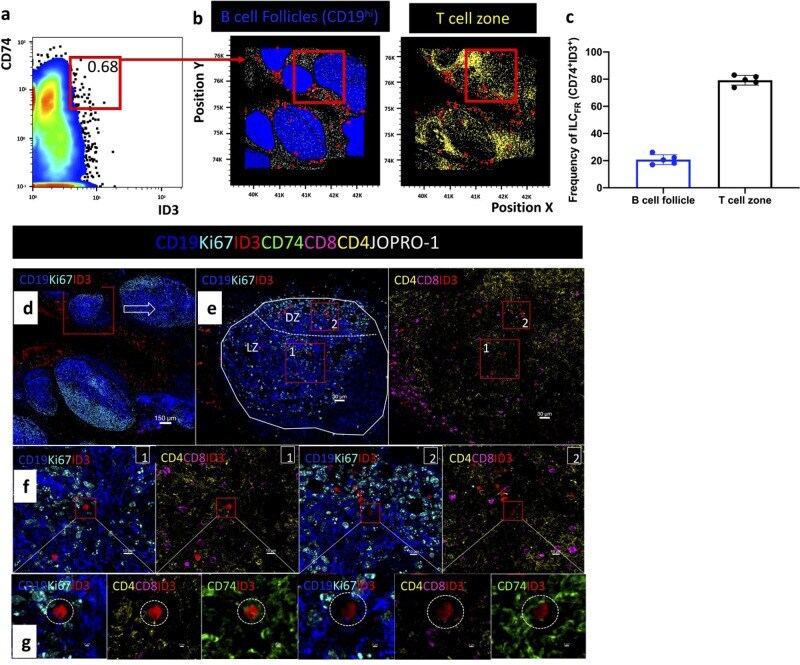
- Fig. 3 Distribution of CD74 + ID3 + ILC FR in human tonsillar B cell follicles. a Histocytometry analysis showing the frequency of CD19- CD8- CD4- CD74+ ID3+ ILC FR in a tonsillar tissue section (tonsil #1) and b Overlay showing the distribution of CD74+ ID3+ ILC FR (red dots) with respect to CD19 + B cells (dark blue dots) and CD4 + T cells (yellow dots) in the same tonsil. c Bar graph summarizing the frequencies of intra- and extra- follicular ILC FR (CD19- CD8- CD4-CD74+ ID3+) as a frequency of total ILC FR in five tonsils. d Confocal images showing the tonsil area imaged, distribution of B cell follicles as denoted by CD19 (dark blue) and Ki67 (cyan) as well as ID3 positive cells (red). e Close up of a B cell follicle. Dotted lines demarcate the area of the follicle (LZ) as well as the dark zone (DZ) as defined by the density of Ki67 staining (cyan). B cells are shown in blue (CD19 + ), proliferating cells in cyan (Ki67 + ), ID3 in red, CD4 in yellow and CD8 in magenta. f Zoomed in details of the red rectangular enclosures shown in ( e ). The location of a CD74 + (green) ID3 + (red) cell is shown with respect to the positioning of CD19 + (blue) Ki67 (cyan) cells or CD8 + (magenta) and CD4 + (yellow) lymphoid cells. g Zoomed in close-ups confirming the positioning and phenotype of ILC FR (CD19-CD4- CD8- CD74+ ID3+). Images were acquired at x40 (NA 1.3) with no zoom. Images shown are sequential digital magnifications of 150 um ( d ), 30 um ( e ), 10 um ( f ), and 2 um ( g )