 Explore
 Validate
 Learn
Learn Flow cytometry
Antibody data
- Antibody Data
- Antigen structure
- References [0]
- Comments [0]
- Validations
- Flow cytometry [3]
- Other assay [2]
Submit
Validation data
Reference
Comment
Report error
- Product number
- MHCD0804 - Provider product page

- Provider
- Invitrogen Antibodies
- Product name
- CD8 Monoclonal Antibody (3B5), PE
- Antibody type
- Monoclonal
- Antigen
- Other
- Description
- R-phycoerythrin (PE) is a stable and highly soluble phycobiliprotein which provides maximal absorbance and fluorescence without susceptibility to internal or external fluorescence quenching, thus providing an exceptional quantum yields and molar extinction coefficients.
- Reactivity
- Human
- Host
- Mouse
- Conjugate
- Yellow dye
- Isotype
- IgG
- Antibody clone number
- 3B5
- Vial size
- 500 µL
- Storage
- 4° C, store in dark
No comments: Submit comment
Supportive validation
- Submitted by
- Invitrogen Antibodies (provider)
- Main image
- Experimental details
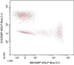
- Dual color flow cytometry dot plot showing mouse anti human CD8 RPE vs. mouse anti human CD4 biotin followed by streptavidin RPE-Alexa Fluor® 610. Human mononuclear cells were stained with mouse anti human CD4 biotin (Product # MHCD0415), washed, stained with streptavidin RPE-Alexa Fluor® 610 (Product # S-20982) and mouse anti human CD8 RPE (Product # MHCD0804), washed, and analyzed on a flow cytometer with a 488 nm excitation source.
- Conjugate
- Yellow dye

- Submitted by
- Invitrogen Antibodies (provider)
- Main image

- Experimental details
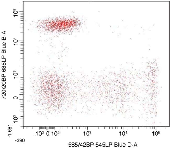
- An antibody complex was made by using the Zenon® Alexa Fluor® 680-R-Phycoerythrin Mouse IgG1 Labeling Kit (Product # Z-25022) and mouse anti human CD4. Human mononuclear cells were stained with mouse anti human CD8 RPE (Product # MHCD0804) and the labled anti-CD4 antibody complex and analyzed by flow cytometry using a 488 nm Laser.
- Conjugate
- Yellow dye
- Submitted by
- Invitrogen Antibodies (provider)
- Main image

- Experimental details
- Human peripheral blood lymphocytes were stained using R-PE of anti-human CD8 monoclonal antibody (clone 3B5). The negative control profiles represent unstained cells.
- Conjugate
- Yellow dye

Supportive validation
- Submitted by
- Invitrogen Antibodies (provider)
- Main image

- Experimental details
- NULL
- Conjugate
- Yellow dye
- Submitted by
- Invitrogen Antibodies (provider)
- Main image

- Experimental details
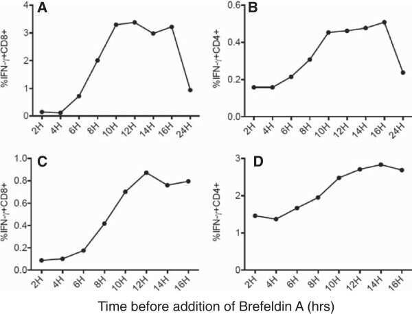
- Figure 4 Antigen-specific CD8 + and CD4 + T cells response following stimulation by DRibbles or cell lysates. (A) The ubiquitinated proteins and pp65 proteins contained in the cell lysate and DRibbles were examined by western blot. Lysates and DRibbles extracted from cell lines treated with bortezomib contained more ubiquitinated proteins than those from non-treated cells (left). The pp65 protein was detected in the lysates and DRibbles extracted from the UbiLT3 pp65 cell line. The pp65 DRibbles collected from the bortezomib-treated UbiLT3 pp65 cell line contained more 148Kd pp65 protein compared with the other groups (right). Data are representative of 3 independent experiments. (B,C) PBMCs were stimulated by DRibbles and cell lysates at the following doses: 3ug/ml, 10ug/ml and 25ug/ml. The percentage of IFN-gamma + CD8 + (B) and IFN-gamma + CD4 + cells (C) were calculated by flow cytometry. Percentages of IFN-gamma + T cells are shown as mean +- SEM. Data are representative of results from 3 independent experiments. (D) Dot plots at the antigen dose of 25ug/ml.
- Conjugate
- Yellow dye
