Explore
 Validate
 Learn
Learn Flow cytometry
Antibody data
- Antibody Data
- Antigen structure
- References [8]
- Comments [0]
- Validations
- Flow cytometry [2]
- Other assay [3]
Submit
Validation data
Reference
Comment
Report error
- Product number
- 25-5273-41 - Provider product page

- Provider
- Invitrogen Antibodies
- Product name
- CD8b Monoclonal Antibody (SIDI8BEE), PE-Cyanine7, eBioscience™
- Antibody type
- Monoclonal
- Antigen
- Other
- Description
- Description: The SIDI8BEE monoclonal antibody reacts with human and rhesus macaque CD8 beta. The CD8b chain associates with the CD8a chain to form the CD8ab heterodimer expressed on the surface of a majority of thymocytes, and on peripheral cytotoxic alpha beta TCR T cells. The CD8 receptor complex binds to MHC class I and plays a role in T cell development and activation of mature T cells. Long been thought to exist either as a heterodimer of the alpha and beta chain or as a homodimer of alpha chains, beta chains have now been shown to homodimerize as well. SIDI8BEE can detect the CD8 beta chain in both the CD8ab heterodimer and the CD8bb homodimer. SIDI8BEE is cross-reactive to human and rhesus macaque CD8b. Other non-human primate species have not been tested. Applications Reported: This SIDI8BEE antibody has been reported for use in flow cytometric analysis. Applications Tested: This SIDI8BEE antibody has been pre-titrated and tested by flow cytometric analysis of normal human peripheral blood cells. This can be used at 5 µL (0.25 µg) per test. A test is defined as the amount (µg) of antibody that will stain a cell sample in a final volume of 100 µL. Cell number should be determined empirically but can range from 10^5 to 10^8 cells/test. Light sensitivity: This tandem dye is sensitive photo-induced oxidation. Please protect this vial and stained samples from light. Fixation: Samples can be stored in IC Fixation Buffer (Product # 00-8222) (100 µL cell sample + 100 µL IC Fixation Buffer) or 1-step Fix/Lyse Solution (Product # 00-5333) for up to 3 days in the dark at 4°C with minimal impact on brightness and FRET efficiency/compensation. Some generalizations regarding fluorophore performance after fixation can be made, but clone specific performance should be determined empirically. Excitation: 488-561 nm; Emission: 775 nm; Laser: Blue Laser, Green Laser, Yellow-Green Laser. Filtration: 0.2 µm post-manufacturing filtered.
- Reactivity
- Human
- Host
- Mouse
- Isotype
- IgG
- Antibody clone number
- SIDI8BEE
- Vial size
- 25 Tests
- Concentration
- 5 µL/Test
- Storage
- 4°C, store in dark, DO NOT FREEZE!
Submitted references Impact of Early ARV Initiation on Relative Proportions of Effector and Regulatory CD8 T Cell in Mesenteric Lymph Nodes and Peripheral Blood During Acute SIV Infection of Rhesus Macaques.
In vitro OP9-DL1 co-culture and subsequent maturation in the presence of IL-21 generates tumor antigen-specific T cells with a favorable less-differentiated phenotype and enhanced functionality.
HIV Infection and Persistence in Pulmonary Mucosal Double Negative T Cells In Vivo.
NKp46-expressing human gut-resident intraepithelial Vδ1 T cell subpopulation exhibits high antitumor activity against colorectal cancer.
Targeted Disruption of HLA Genes via CRISPR-Cas9 Generates iPSCs with Enhanced Immune Compatibility.
Antigen receptor-redirected T cells derived from hematopoietic precursor cells lack expression of the endogenous TCR/CD3 receptor and exhibit specific antitumor capacities.
CD103+ intraepithelial T cells in high-grade serous ovarian cancer are phenotypically diverse TCRαβ+ CD8αβ+ T cells that can be targeted for cancer immunotherapy.
GATA3 induces human T-cell commitment by restraining Notch activity and repressing NK-cell fate.
Yero A, Farnos O, Clain J, Zghidi-Abouzid O, Rabezanahary H, Racine G, Estaquier J, Jenabian MA
Journal of virology 2022 Apr 13;96(7):e0025522
Journal of virology 2022 Apr 13;96(7):e0025522
In vitro OP9-DL1 co-culture and subsequent maturation in the presence of IL-21 generates tumor antigen-specific T cells with a favorable less-differentiated phenotype and enhanced functionality.
Bonte S, de Munter S, Billiet L, Goetgeluk G, Ingels J, Jansen H, Pille M, de Cock L, Weening K, Taghon T, Leclercq G, Vandekerckhove B, Kerre T
Oncoimmunology 2021;10(1):1954800
Oncoimmunology 2021;10(1):1954800
HIV Infection and Persistence in Pulmonary Mucosal Double Negative T Cells In Vivo.
Meziane O, Salahuddin S, Pham TNQ, Farnos O, Pagliuzza A, Olivenstein R, Thomson E, Alexandrova Y, Orlova M, Schurr E, Ancuta P, Haddad É, Chomont N, Cohen EA, Jenabian MA, Costiniuk CT
Journal of virology 2020 Nov 23;94(24)
Journal of virology 2020 Nov 23;94(24)
NKp46-expressing human gut-resident intraepithelial Vδ1 T cell subpopulation exhibits high antitumor activity against colorectal cancer.
Mikulak J, Oriolo F, Bruni E, Roberto A, Colombo FS, Villa A, Bosticardo M, Bortolomai I, Lo Presti E, Meraviglia S, Dieli F, Vetrano S, Danese S, Della Bella S, Carvello MM, Sacchi M, Cugini G, Colombo G, Klinger M, Spaggiari P, Roncalli M, Prinz I, Ravens S, di Lorenzo B, Marcenaro E, Silva-Santos B, Spinelli A, Mavilio D
JCI insight 2019 Dec 19;4(24)
JCI insight 2019 Dec 19;4(24)
Targeted Disruption of HLA Genes via CRISPR-Cas9 Generates iPSCs with Enhanced Immune Compatibility.
Xu H, Wang B, Ono M, Kagita A, Fujii K, Sasakawa N, Ueda T, Gee P, Nishikawa M, Nomura M, Kitaoka F, Takahashi T, Okita K, Yoshida Y, Kaneko S, Hotta A
Cell stem cell 2019 Apr 4;24(4):566-578.e7
Cell stem cell 2019 Apr 4;24(4):566-578.e7
Antigen receptor-redirected T cells derived from hematopoietic precursor cells lack expression of the endogenous TCR/CD3 receptor and exhibit specific antitumor capacities.
Van Caeneghem Y, De Munter S, Tieppo P, Goetgeluk G, Weening K, Verstichel G, Bonte S, Taghon T, Leclercq G, Kerre T, Debets R, Vermijlen D, Abken H, Vandekerckhove B
Oncoimmunology 2017;6(3):e1283460
Oncoimmunology 2017;6(3):e1283460
CD103+ intraepithelial T cells in high-grade serous ovarian cancer are phenotypically diverse TCRαβ+ CD8αβ+ T cells that can be targeted for cancer immunotherapy.
Komdeur FL, Wouters MC, Workel HH, Tijans AM, Terwindt AL, Brunekreeft KL, Plat A, Klip HG, Eggink FA, Leffers N, Helfrich W, Samplonius DF, Bremer E, Wisman GB, Daemen T, Duiker EW, Hollema H, Nijman HW, de Bruyn M
Oncotarget 2016 Nov 15;7(46):75130-75144
Oncotarget 2016 Nov 15;7(46):75130-75144
GATA3 induces human T-cell commitment by restraining Notch activity and repressing NK-cell fate.
Van de Walle I, Dolens AC, Durinck K, De Mulder K, Van Loocke W, Damle S, Waegemans E, De Medts J, Velghe I, De Smedt M, Vandekerckhove B, Kerre T, Plum J, Leclercq G, Rothenberg EV, Van Vlierberghe P, Speleman F, Taghon T
Nature communications 2016 Apr 6;7:11171
Nature communications 2016 Apr 6;7:11171
No comments: Submit comment
Supportive validation
- Submitted by
- Invitrogen Antibodies (provider)
- Main image
- Experimental details
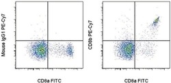
- Staining of normal human peripheral blood cells with Anti-Human CD8a FITC (Product # 11-0087-42) and Mouse IgG1 K Isotype Control PE-Cyanine7 (Product # 25-4714-80) (left) or Anti-Human/Non-Human Primate CD8b PE-Cyanine7 (right). Cells in the lymphocyte gate were used for analysis.
- Submitted by
- Invitrogen Antibodies (provider)
- Main image

- Experimental details
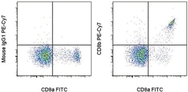
- Staining of normal human peripheral blood cells with Anti-Human CD8a FITC (Product # 11-0087-42) and Mouse IgG1 K Isotype Control PE-Cyanine7 (Product # 25-4714-80) (left) or Anti-Human/Non-Human Primate CD8b PE-Cyanine7 (right). Cells in the lymphocyte gate were used for analysis.

Supportive validation
- Submitted by
- Invitrogen Antibodies (provider)
- Main image
- Experimental details
- NULL
- Submitted by
- Invitrogen Antibodies (provider)
- Main image
- Experimental details
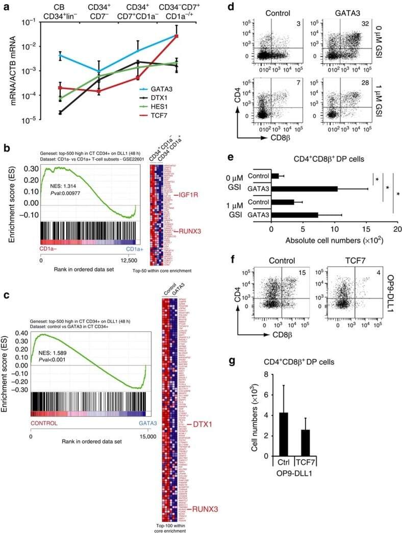
- Figure 4 GATA3 restrains Notch activation at the T-lineage commitment stage. ( a ) Quantitative PCR of GATA3 , DTX1 , HES1 and TCF7 expression in different stages of in vitro generated T cell precursors from CB CD34 + lin - HPCs after 7 days of OP9-DLL4 coculture. Data shows the average expression in 3-4 independent samples on a log scale and erros bars indicate s.e.m. ( b , c ) GSEA shows a significant enrichment of the top 500 Notch-dependent genes in human CD34 + thymocytes in the set of genes higher expressed in ( b ) uncommitted CD34 + CD1a - versus CD34 + CD1a + committed T-cell precursors 50 , and genes expressed higher in ( c ) control versus GATA3-transduced CD34 + thymocytes as determined by microarray after 48 h of transduction. ( d ) Flow cytometry analysis of control and GATA3 -transduced CD34 + CD1 - uncommitted thymocytes in OP9-DLL1 cocultures with addition of 0 or 1 muM GSI and in the presence of IL7, SCF and FLT3L, showing the development of CD4 + CD8beta + DP thymocytes after 6 days of coculture. ( e ) Graph show absolute number of CD4 + CD8beta + DP thymocytes, generated in corresponding cultures shown in d . Data shows the average of four independent experiments and errors bars show s.e.m. * P
- Submitted by
- Invitrogen Antibodies (provider)
- Main image
- Experimental details
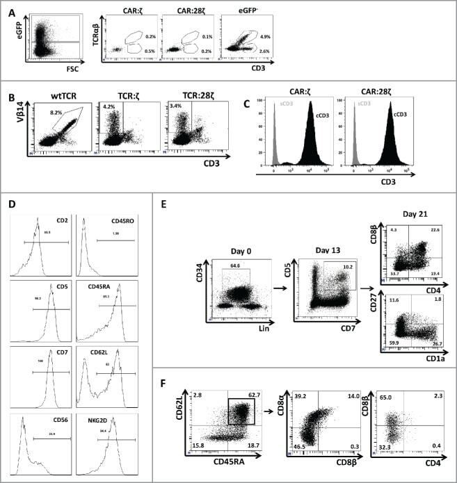
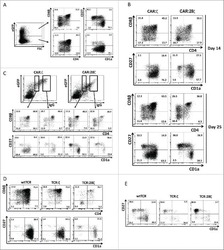
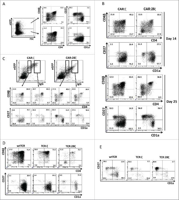
- Figure 3. Phenotype and endogenous TCR expression of CD34 + HPC-derived transgenic AR + T cells. Flow cytometric analysis of the AR-transgenic T cells. (A) CAR-transgenic GFP + cells of cultures transduced to express either the CAR:zeta or the CAR:28zeta were analyzed on day 26 of OP9-DL1 culture for CD3 and TCRalphabeta expression. As a control, GFP - cells are shown from the OP9-DL1 culture transduced to express the CAR:zeta ( N = 5). (B) Dot plots show CD3 expression of cells from the OP9-DL1 cultures transgenic for the wtTCR, TCR:zeta and TCR:28zeta. Vbeta14 staining is used to mark transgene expression, as no GFP is expressed by the transgenic cells ( N = 5). (C) Surface and cytoplasmic staining for CD3 of in vitro generated mature T cells that were expanded for one cycle on feeder cells in the presence of cytokines. (D) Expression of various membrane markers by the CD27 + CD1a - mature T cells at the end of OP9-DL1 culture (46 d) ( N = 2). (E) Day 0: fresh cord blood after MACS CD34 enrichment sorted using the sorting window shown. Day 13: cord blood cells cultured on OP9-DL1 were sorted for CD5 CD7 double positive cells, using the indicated sorting window. The cells were then transduced to express CAR:28zeta and further differentiated on OP9-DL1 feeder layer. Day 21: analysis of the transgenic GFP + cultured cells for DP cells and CD27 + CD1a - mature cells. (F) Flow cytometric analysis of GFP + CAR:28zeta-transgenic cultures, gated on GFP + CD27 + CD1a