Explore
 Validate
 Learn
Learn Western blot
 Immunocytochemistry
Antibody data
- Antibody Data
- Antigen structure
- References [8]
- Comments [0]
- Validations
- Immunocytochemistry [8]
- Immunohistochemistry [1]
- Other assay [4]
Submit
Validation data
Reference
Comment
Report error
- Product number
- PA1-092 - Provider product page

- Provider
- Invitrogen Antibodies
- Product name
- Cdc42 Polyclonal Antibody
- Antibody type
- Polyclonal
- Antigen
- Recombinant full-length protein
- Reactivity
- Human, Mouse, Rat
- Host
- Rabbit
- Isotype
- IgG
- Vial size
- 100 μg
- Concentration
- 1 mg/mL
- Storage
- -20°C, Avoid Freeze/Thaw Cycles
Submitted references A nuclear function for an oncogenic microRNA as a modulator of snRNA and splicing.
Cdc42 negatively regulates endocytosis during apical membrane maintenance in live animals.
Phenotypic characterisation of breast cancer: the role of CDC42.
Endocytic Pathways Used by Andes Virus to Enter Primary Human Lung Endothelial Cells.
The NarE protein of Neisseria gonorrhoeae catalyzes ADP-ribosylation of several ADP-ribose acceptors despite an N-terminal deletion.
The localisation of the apical Par/Cdc42 polarity module is specifically affected in microvillus inclusion disease.
MicroRNA-27a Inhibits Cell Migration and Invasion of Fibroblast-Like Synoviocytes by Targeting Follistatin-Like Protein 1 in Rheumatoid Arthritis.
14-3-3ε and ζ regulate neurogenesis and differentiation of neuronal progenitor cells in the developing brain.
El Fatimy R, Zhang Y, Deforzh E, Ramadas M, Saravanan H, Wei Z, Rabinovsky R, Teplyuk NM, Uhlmann EJ, Krichevsky AM
Molecular cancer 2022 Jan 15;21(1):17
Molecular cancer 2022 Jan 15;21(1):17
Cdc42 negatively regulates endocytosis during apical membrane maintenance in live animals.
Shitara A, Malec L, Ebrahim S, Chen D, Bleck C, Hoffman MP, Weigert R
Molecular biology of the cell 2019 Feb 1;30(3):324-332
Molecular biology of the cell 2019 Feb 1;30(3):324-332
Phenotypic characterisation of breast cancer: the role of CDC42.
Chrysanthou E, Gorringe KL, Joseph C, Craze M, Nolan CC, Diez-Rodriguez M, Green AR, Rakha EA, Ellis IO, Mukherjee A
Breast cancer research and treatment 2017 Jul;164(2):317-325
Breast cancer research and treatment 2017 Jul;164(2):317-325
Endocytic Pathways Used by Andes Virus to Enter Primary Human Lung Endothelial Cells.
Chiang CF, Flint M, Lin JS, Spiropoulou CF
PloS one 2016;11(10):e0164768
PloS one 2016;11(10):e0164768
The NarE protein of Neisseria gonorrhoeae catalyzes ADP-ribosylation of several ADP-ribose acceptors despite an N-terminal deletion.
Rodas PI, Álamos-Musre AS, Álvarez FP, Escobar A, Tapia CV, Osorio E, Otero C, Calderón IL, Fuentes JA, Gil F, Paredes-Sabja D, Christodoulides M
FEMS microbiology letters 2016 Sep;363(17)
FEMS microbiology letters 2016 Sep;363(17)
The localisation of the apical Par/Cdc42 polarity module is specifically affected in microvillus inclusion disease.
Michaux G, Massey-Harroche D, Nicolle O, Rabant M, Brousse N, Goulet O, Le Bivic A, Ruemmele FM
Biology of the cell 2016 Jan;108(1):19-28
Biology of the cell 2016 Jan;108(1):19-28
MicroRNA-27a Inhibits Cell Migration and Invasion of Fibroblast-Like Synoviocytes by Targeting Follistatin-Like Protein 1 in Rheumatoid Arthritis.
Shi DL, Shi GR, Xie J, Du XZ, Yang H
Molecules and cells 2016 Aug 31;39(8):611-8
Molecules and cells 2016 Aug 31;39(8):611-8
14-3-3ε and ζ regulate neurogenesis and differentiation of neuronal progenitor cells in the developing brain.
Toyo-oka K, Wachi T, Hunt RF, Baraban SC, Taya S, Ramshaw H, Kaibuchi K, Schwarz QP, Lopez AF, Wynshaw-Boris A
The Journal of neuroscience : the official journal of the Society for Neuroscience 2014 Sep 3;34(36):12168-81
The Journal of neuroscience : the official journal of the Society for Neuroscience 2014 Sep 3;34(36):12168-81
No comments: Submit comment
Supportive validation
- Submitted by
- Invitrogen Antibodies (provider)
- Main image

- Experimental details
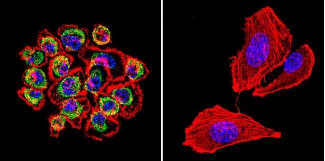
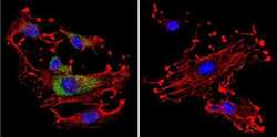
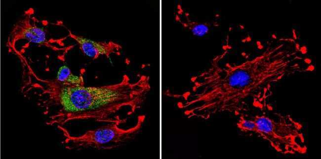
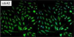
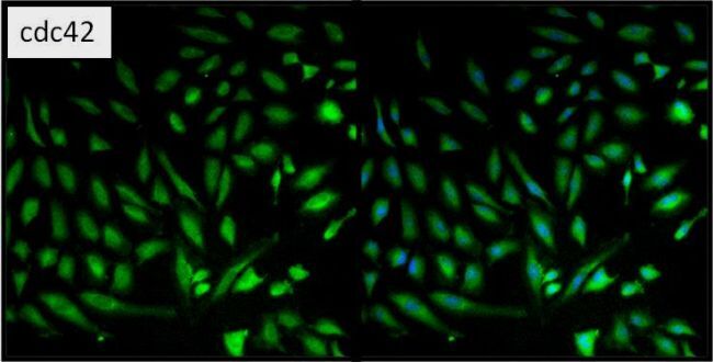
- Immunofluorescent analysis of cdc42 in A431 Cells. Cells were grown on chamber slides and fixed with formaldehyde prior to staining. Cells were probed without (control) or with a cdc42 polyclonal antibody (Product # PA1-092) at a dilution of 1:20 overnight at 4 C, washed with PBS and incubated with a DyLight-488 conjugated secondary antibody (Product # 35552). cdc42 staining (green), F-Actin staining with Phalloidin (red) and nuclei with DAPI (blue) is shown. Images were taken at 60X magnification.
- Submitted by
- Invitrogen Antibodies (provider)
- Main image
- Experimental details
- Immunofluorescent analysis of cdc42 in C6 Cells. Cells were grown on chamber slides and fixed with formaldehyde prior to staining. Cells were probed without (control) or with a cdc42 polyclonal antibody (Product # PA1-092) at a dilution of 1:20 overnight at 4 C, washed with PBS and incubated with a DyLight-488 conjugated secondary antibody (Product # 35552). cdc42 staining (green), F-Actin staining with Phalloidin (red) and nuclei with DAPI (blue) is shown. Images were taken at 60X magnification.
- Submitted by
- Invitrogen Antibodies (provider)
- Main image
- Experimental details
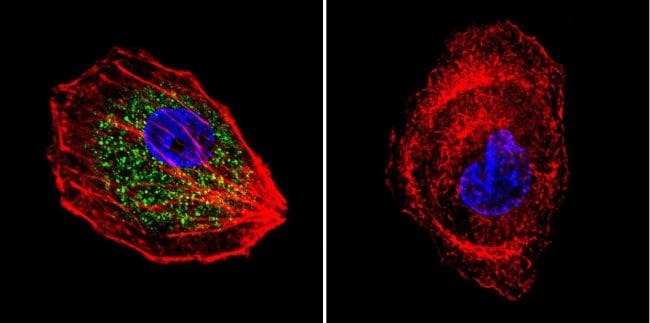
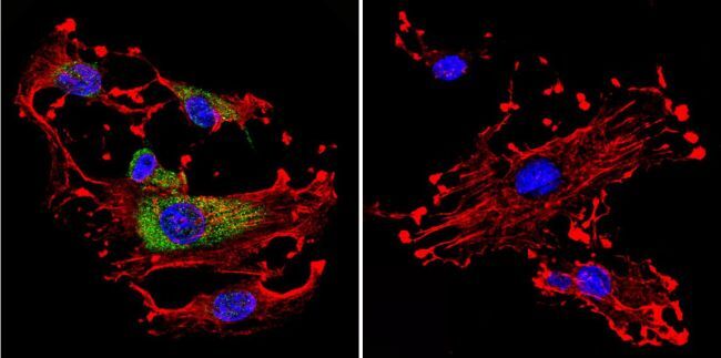
- Immunofluorescent analysis of cdc42 (green) in HeLa cells. Formalin fixed cells were permeabilized with 0.1% Triton X-100 in TBS for 10 minutes at room temperature and blocked with 5% normal goat serum (Product # 31873) for 15 minutes at room temperature. Cells were probed with a cdc42 polyclonal antibody (Product # PA1-092) at a dilution of 1:100 for at least 1 hour at room temperature, washed with PBS, and incubated with DyLight 488 goat anti-rabbit IgG secondary antibody (Product # 35552) at a dilution of 1:400 for 30 minutes at room temperature. Nuclei (blue) were stained with Hoechst 33342 dye (Product # 62249). Images were taken on a Thermo Scientific ArrayScan at 20X magnification.

- Submitted by
- Invitrogen Antibodies (provider)
- Main image

- Experimental details
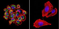
- Immunofluorescent analysis of cdc42 in HepG2 Cells. Cells were grown on chamber slides and fixed with formaldehyde prior to staining. Cells were probed without (control) or with a cdc42 polyclonal antibody (Product # PA1-092) at a dilution of 1:200 overnight at 4 C, washed with PBS and incubated with a DyLight-488 conjugated secondary antibody (Product # 35552). cdc42 staining (green), F-Actin staining with Phalloidin (red) and nuclei with DAPI (blue) is shown. Images were taken at 60X magnification.
- Submitted by
- Invitrogen Antibodies (provider)
- Main image
- Experimental details
- Immunofluorescent analysis of cdc42 in A431 Cells. Cells were grown on chamber slides and fixed with formaldehyde prior to staining. Cells were probed without (control) or with a cdc42 polyclonal antibody (Product # PA1-092) at a dilution of 1:20 overnight at 4 C, washed with PBS and incubated with a DyLight-488 conjugated secondary antibody (Product # 35552). cdc42 staining (green), F-Actin staining with Phalloidin (red) and nuclei with DAPI (blue) is shown. Images were taken at 60X magnification.

- Submitted by
- Invitrogen Antibodies (provider)
- Main image
- Experimental details
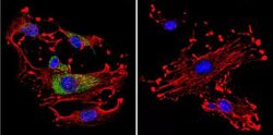
- Immunofluorescent analysis of cdc42 in C6 Cells. Cells were grown on chamber slides and fixed with formaldehyde prior to staining. Cells were probed without (control) or with a cdc42 polyclonal antibody (Product # PA1-092) at a dilution of 1:20 overnight at 4 C, washed with PBS and incubated with a DyLight-488 conjugated secondary antibody (Product # 35552). cdc42 staining (green), F-Actin staining with Phalloidin (red) and nuclei with DAPI (blue) is shown. Images were taken at 60X magnification.
- Submitted by
- Invitrogen Antibodies (provider)
- Main image

- Experimental details
- Immunofluorescent analysis of cdc42 in HepG2 Cells. Cells were grown on chamber slides and fixed with formaldehyde prior to staining. Cells were probed without (control) or with a cdc42 polyclonal antibody (Product # PA1-092) at a dilution of 1:200 overnight at 4 C, washed with PBS and incubated with a DyLight-488 conjugated secondary antibody (Product # 35552). cdc42 staining (green), F-Actin staining with Phalloidin (red) and nuclei with DAPI (blue) is shown. Images were taken at 60X magnification.

- Submitted by
- Invitrogen Antibodies (provider)
- Main image

- Experimental details
- Immunofluorescent analysis of cdc42 (green) in HeLa cells. Formalin fixed cells were permeabilized with 0.1% Triton X-100 in TBS for 10 minutes at room temperature and blocked with 5% normal goat serum (Product # 31873) for 15 minutes at room temperature. Cells were probed with a cdc42 polyclonal antibody (Product # PA1-092) at a dilution of 1:100 for at least 1 hour at room temperature, washed with PBS, and incubated with DyLight 488 goat anti-rabbit IgG secondary antibody (Product # 35552) at a dilution of 1:400 for 30 minutes at room temperature. Nuclei (blue) were stained with Hoechst 33342 dye (Product # 62249). Images were taken on a Thermo Scientific ArrayScan at 20X magnification.
Supportive validation
- Submitted by
- Invitrogen Antibodies (provider)
- Main image
- Experimental details
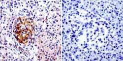
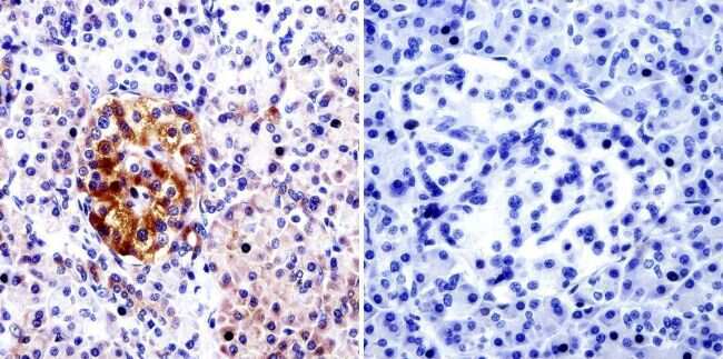
- Immunohistochemistry was performed on normal biopsies of deparaffinized human pancreas tissue. To expose target proteins, heat induced antigen retrieval was performed using 10mM sodium citrate (pH6.0) buffer, microwaved for 8-15 minutes. Following antigen retrieval tissues were blocked in 3% BSA-PBS for 30 minutes at room temperature. Tissues were then probed at a dilution of 1:20 with a Rabbit Polyclonal Antibody recognizing cdc42 (Product # PA1-092) or without primary antibody (negative control) overnight at 4°C in a humidified chamber. Tissues were washed extensively with PBST and endogenous peroxidase activity was quenched with a peroxidase suppressor. Detection was performed using a biotin-conjugated secondary antibody and SA-HRP, followed by colorimetric detection using DAB. Tissues were counterstained with hematoxylin and prepped for mounting.
Supportive validation
- Submitted by
- Invitrogen Antibodies (provider)
- Main image
- Experimental details
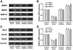
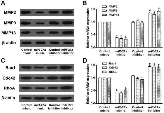
- Fig. 3. Effects of miR-27a on the expression of migration and invasion-related proteins in RA-FLS. RA-FLS were transfected with miR-27a mimic or miR-27a inhibitor. (A) The protein and mRNA expression of MMP2, MMP9, and MMP13 in RA-FLS was detected by western blot and qRT-PCR assay. (B) The protein and mRNA expression of Rac1, Cdc42, and RhoA in RA-FLS was detected by western blot and qRT-PCR assay. * p < 0.05, versus control mimic group. # p < 0.05, versus control inhibitor group.
- Submitted by
- Invitrogen Antibodies (provider)
- Main image
- Experimental details
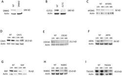
- Fig 2 Knockdown of selected genes required for ANDV infection. Western blots of indicated proteins after knockdown with gene-specific siRNA against (A) dynamin 2 (DNM2); (B) clathrin heavy chain (CLTC); (C) AP2M1; (D) caveolin 1 (CAV1); (E) CDC42; (F) ARF6; (G) NSF; (H) RAB5C; or (I) TSG101; or non-targeting siRNA transfection control (NT). siRNAs were transfected into HMVEC-L at the concentration of 100 nM for 48 h (see Table 1 for more information regarding these genes). The cells were then infected with ANDV (MOI = 0.5) for 24 h or 48 h. Western blots were performed post infection to ensure knockdown of the specific protein expression. Molecular weight of each specific protein is indicated on the right side of each panel. The blots were also probed with beta-actin specific antibody as the gel-loading control. (A) and (B) show only the results collected at 48 h.
- Submitted by
- Invitrogen Antibodies (provider)
- Main image
- Experimental details
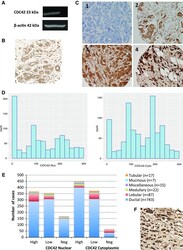
- Fig. 1 Expression of CDC42 in breast cancer. a Western Blotting analysis using anti-CDC42 Polyclonal Antibody (PA1-092) showing band at the expected size (23 kDa). b CDC42 expression in terminal duct lobular units using IHC. c CDC42 expression in TMA cores using IHC. Intensity levels of staining are shown: 1 negative, 2 weak, 3 moderate and 4 strong expression (x20 magnification). d Histograms of H-scores for nuclear and cytoplasmic staining. e Distribution of cases across different histological subtypes, showing the increased proportion of lobular cases with high CDC42 nuclear staining but low or negative cytoplasmic staining. f Example of lobular carcinoma showing strong nuclear staining
- Submitted by
- Invitrogen Antibodies (provider)
- Main image
- Experimental details
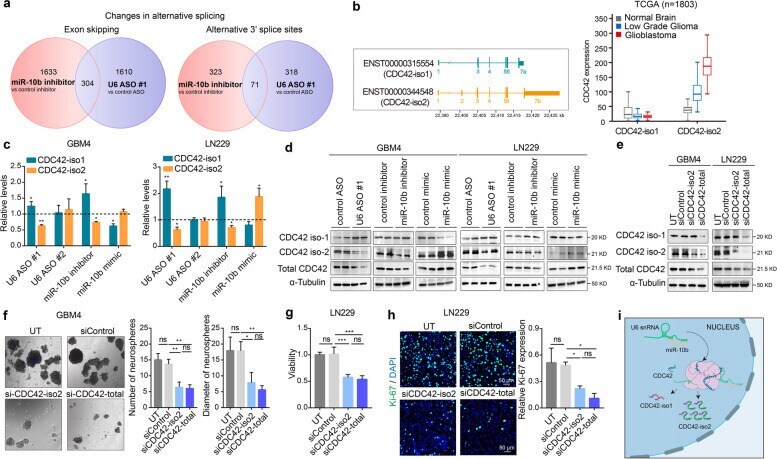
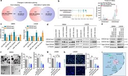
- Fig. 5 miR-10b regulates alternative splicing of CDC42, thereby controlling its total levels via U6 regulation. a Alternative splicing events induced by miR-10b inhibitor and U6 ASO #1, as determined by RNAseq ( n = 3). Venn diagrams indicate the numbers of exon skipping (SE) and alternative 3' splice sites (A3SS) modulation, in the indicated conditions. b Schematic illustration of two major CDC42 mRNA isoforms, with alternative exons encoding alternative 5' and 3' UTRs (left panel). Expression analysis of CDC42 isoforms in the normal brain ( n = 1141), low grade glioma (LGG) ( n = 509) and GBM ( n = 153) in TCGA database demonstrates that ENST00000344548 (CDC42-iso2) is the pathologic variant associated with glioma progression, whereas ENST00000315554 (CDC42-iso1) is present at similarly low levels in the normal brain, LGG, and GBM (right panel). c qRT-PCR analysis of CDC42-iso1 or iso2 mRNAs in glioma cells and GSCs transfected with either U6 ASOs, or miR-10b inhibitor or mimic. The results are expressed as the fold-changes relative to the corresponding control groups (mean +- SD, n = 3). P values were calculated using two-tail unpaired t-test. d Western blot analysis of the indicated CDC42 forms in the cells transfected with either U6 ASO, or miR-10b inhibitor or mimic. e Western blot analysis of the cells transfected with either selective siRNA targeting iso2 (siCDC42-iso2) or both CDC42 isoforms (siCDC42-total) demonstrates that KD of iso2 is sufficient for reducing tota