Explore
 Validate
 Learn
Learn Western blot
 Chromatin Immunoprecipitation
Antibody data
- Antibody Data
- Antigen structure
- References [0]
- Comments [0]
- Validations
- Western blot [3]
- Immunocytochemistry [1]
- Immunohistochemistry [14]
- Other assay [1]
Submit
Validation data
Reference
Comment
Report error
- Product number
- UM570011 - Provider product page

- Provider
- Invitrogen Antibodies
- Product name
- ERCC1 Monoclonal Antibody (2E12), UltraMAB™
- Antibody type
- Monoclonal
- Antigen
- Recombinant full-length protein
- Reactivity
- Human, Rat
- Host
- Mouse
- Isotype
- IgG
- Antibody clone number
- 2E12
- Vial size
- 30 µL
- Concentration
- 0.5-1.0 mg/mL
- Storage
- -20° C, Avoid Freeze/Thaw Cycles
No comments: Submit comment
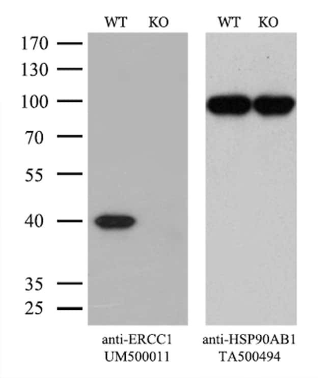
Supportive validation
- Submitted by
- Invitrogen Antibodies (provider)
- Main image
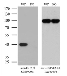
- Experimental details
- Equivalent amounts of cell lysates (10 µg per lane) of wild-type Hela cells (WT, Cat# LC810HELA) and ERCC1-Knockout Hela cells (KO, Cat# LC810072) were separated by SDS-PAGE and immunoblotted with anti-ERCC1 monoclonal antibody UM500011. Then the blotted membrane was stripped and reprobed with anti-HSP90AB1 antibody (TA500494) as a loading control. (1:500)
- Submitted by
- Invitrogen Antibodies (provider)
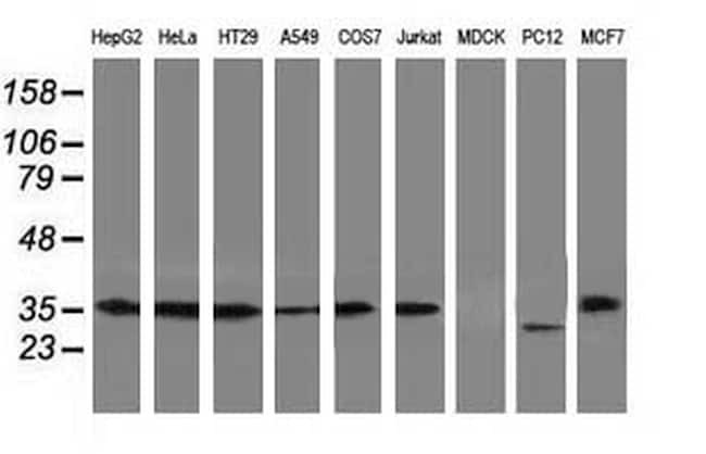
- Main image

- Experimental details
- Western blot analysis of extracts (35 µg) from 9 different cell lines by using anti-ERCC1 monoclonal antibody (Clone 2E12).
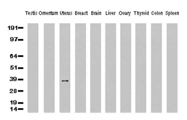
- Submitted by
- Invitrogen Antibodies (provider)
- Main image
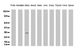
- Experimental details
- Western Blot analysis of 10 different human tissue lysates (10 µg) by using anti-ERCC1 monoclonal antibody (clone 2E12, 1:500)
Supportive validation
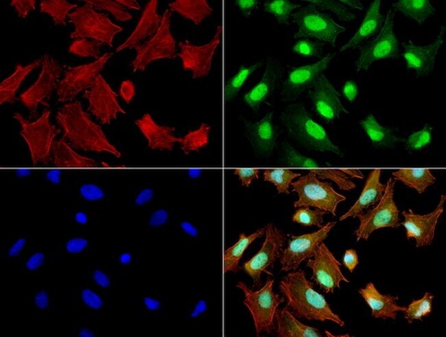
- Submitted by
- Invitrogen Antibodies (provider)
- Main image
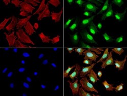
- Experimental details
- Immunofluorescent staining of HeLa cells using ERCC1 mouse monoclonal antibody (UM500011, green). Actin filaments were labeled with TRITC-phalloidin (red), and nuclear with DAPI (blue). The three-color overlay image is located at the bottom-right corner.
Supportive validation
- Submitted by
- Invitrogen Antibodies (provider)
- Main image
- Experimental details
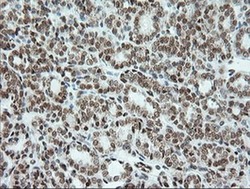
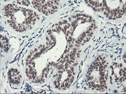
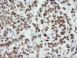
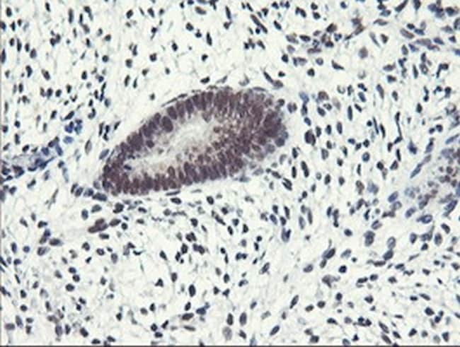
- Immunohistochemical staining of paraffin-embedded Carcinoma of Human thyroid tissue using anti-ERCC1 mouse monoclonal antibody. (Clone 2E12, dilution 1:100; heat-induced epitope retrieval by 10mM citric buffer, pH6.0, 120°C for 3min)
- Submitted by
- Invitrogen Antibodies (provider)
- Main image
- Experimental details
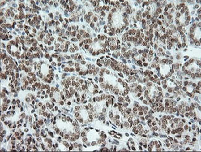
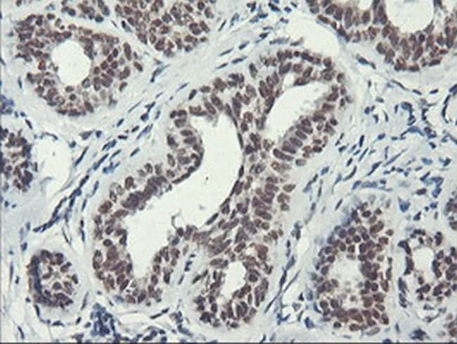
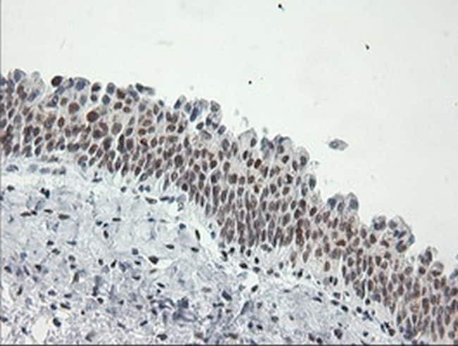
- Immunohistochemical staining of paraffin-embedded Carcinoma of Human bladder tissue using anti-ERCC1 mouse monoclonal antibody. (Clone 2E12, dilution 1:100; heat-induced epitope retrieval by 10mM citric buffer, pH6.0, 120°C for 3min)
- Submitted by
- Invitrogen Antibodies (provider)
- Main image
- Experimental details
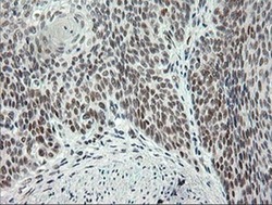
- Immunohistochemical staining of paraffin-embedded human breast tissue using anti-ERCC1 mouse monoclonal antibody. (Clone 2E12, dilution 1:100; heat-induced epitope retrieval by 10mM citric buffer, pH6.0, 120°C for 3min)
- Submitted by
- Invitrogen Antibodies (provider)
- Main image

- Experimental details
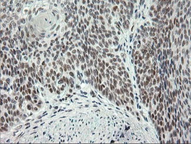
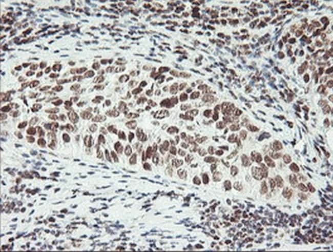
- Immunohistochemical staining of paraffin-embedded Carcinoma of Human lung tissue using anti-ERCC1 mouse monoclonal antibody. (Clone 2E12, dilution 1:100; heat-induced epitope retrieval by 10mM citric buffer, pH6.0, 120°C for 3min)
- Submitted by
- Invitrogen Antibodies (provider)
- Main image

- Experimental details
- Immunohistochemical staining of paraffin-embedded human Ovary tissue using anti-ERCC1 mouse monoclonal antibody. (Clone 2E12, dilution 1:100; heat-induced epitope retrieval by 10mM citric buffer, pH6.0, 120°C for 3min)
- Submitted by
- Invitrogen Antibodies (provider)
- Main image
- Experimental details
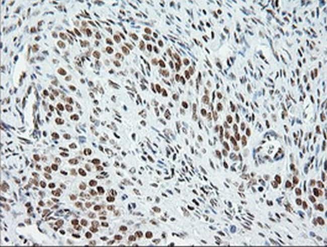
- Immunohistochemical staining of paraffin-embedded Adenocarcinoma of Human ovary tissue using anti-ERCC1 mouse monoclonal antibody. (Clone 2E12, dilution 1:100; heat-induced epitope retrieval by 10mM citric buffer, pH6.0, 120°C for 3min)

- Submitted by
- Invitrogen Antibodies (provider)
- Main image
- Experimental details
- Immunohistochemical staining of paraffin-embedded human pancreas tissue using anti-ERCC1 mouse monoclonal antibody. (Clone 2E12, dilution 1:100; heat-induced epitope retrieval by 10mM citric buffer, pH6.0, 120°C for 3min)

- Submitted by
- Invitrogen Antibodies (provider)
- Main image

- Experimental details
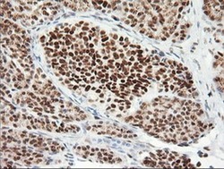
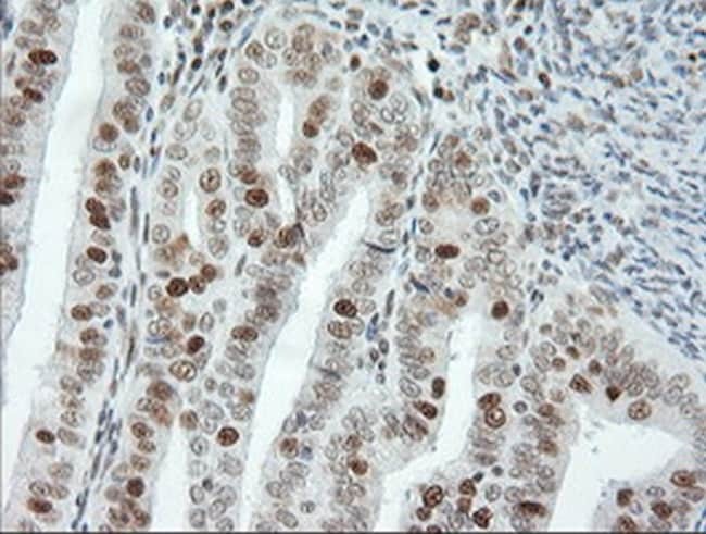
- Immunohistochemical staining of paraffin-embedded human thyroid tissue using anti-ERCC1 mouse monoclonal antibody. (Clone 2E12, dilution 1:100; heat-induced epitope retrieval by 10mM citric buffer, pH6.0, 120°C for 3min)
- Submitted by
- Invitrogen Antibodies (provider)
- Main image
- Experimental details
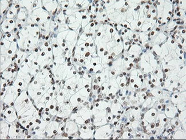
- Immunohistochemical staining of paraffin-embedded human endometrium tissue using anti-ERCC1 mouse monoclonal antibody. (Clone 2E12, dilution 1:100; heat-induced epitope retrieval by 10mM citric buffer, pH6.0, 120°C for 3min)
- Submitted by
- Invitrogen Antibodies (provider)
- Main image

- Experimental details
- Immunohistochemical staining of paraffin-embedded Adenocarcinoma of Human endometrium tissue using anti-ERCC1 mouse monoclonal antibody. (Clone 2E12, dilution 1:100; heat-induced epitope retrieval by 10mM citric buffer, pH6.0, 120°C for 3min)
- Submitted by
- Invitrogen Antibodies (provider)
- Main image

- Experimental details
- Immunohistochemical staining of paraffin-embedded human bladder tissue using anti-ERCC1 mouse monoclonal antibody. (Clone 2E12, dilution 1:100; heat-induced epitope retrieval by 10mM citric buffer, pH6.0, 120°C for 3min)
- Submitted by
- Invitrogen Antibodies (provider)
- Main image

- Experimental details
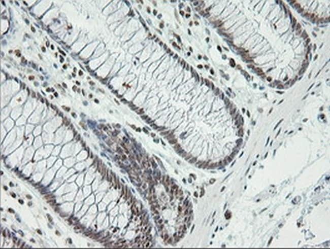
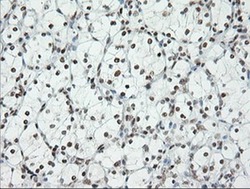
- Immunohistochemical staining of paraffin-embedded human colon tissue using anti-ERCC1 mouse monoclonal antibody. (Clone 2E12, dilution 1:100; heat-induced epitope retrieval by 10mM citric buffer, pH6.0, 120°C for 3min)
- Submitted by
- Invitrogen Antibodies (provider)
- Main image
- Experimental details
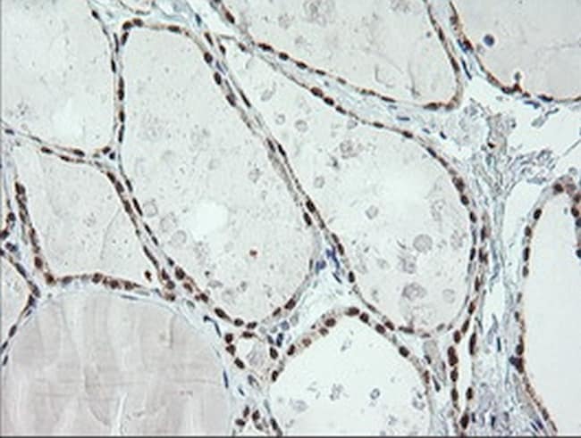
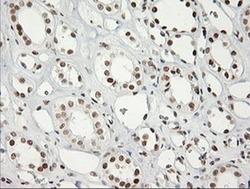
- Immunohistochemical staining of paraffin-embedded human Kidney tissue using anti-ERCC1 mouse monoclonal antibody. (Clone 2E12, dilution 1:100; heat-induced epitope retrieval by 10mM citric buffer, pH6.0, 120°C for 3min)
- Submitted by
- Invitrogen Antibodies (provider)
- Main image
- Experimental details
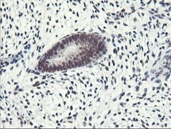
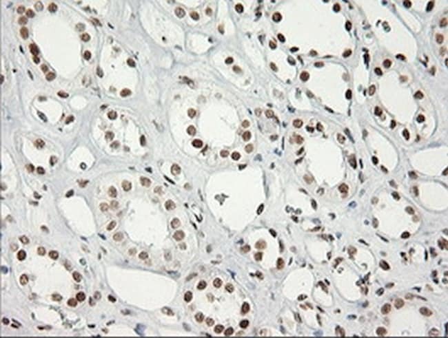
- Immunohistochemical staining of paraffin-embedded Carcinoma of Human kidney tissue using anti-ERCC1 mouse monoclonal antibody. (Clone 2E12, dilution 1:100; heat-induced epitope retrieval by 10mM citric buffer, pH6.0, 120°C for 3min)
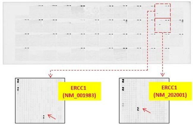
Supportive validation
- Submitted by
- Invitrogen Antibodies (provider)
- Main image
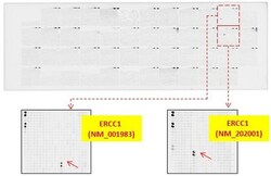
- Experimental details
- OriGene overexpression protein microarray chip was immunostained with UltraMAB anti-ERCC1 mouse monoclonal antibody (Clone 2E12). The positive reactive proteins are highlighted with red arrows in the enlarged subarray. Other positive controls spotted in this subarray are serial dilutions of mouse IgG as controls.