 Explore
 Validate
 Learn
Learn Western blot
 Immunohistochemistry
Antibody data
- Antibody Data
- Antigen structure
- References [3]
- Comments [0]
- Validations
- Immunohistochemistry [1]
Submit
Validation data
Reference
Comment
Report error
- Product number
- AF-273-NA - Provider product page

- Provider
- Novus Biologicals
- Product name
- Goat Polyclonal FGF-9 Antibody
- Antibody type
- Polyclonal
- Description
- Antigen Affinity-purified. Detects human FGF-9 in direct ELISAs and Western blots. In direct ELISAs, less than 1% cross-reactivity with recombinant human (rh) FGF-4, rhFGF-5, rhFGF-6, rhFGF-7, rhFGF-8, rhFGF-10, rhFGF-17, rhFGF-18, and rhFGF-19 is observed.
- Reactivity
- Human
- Host
- Goat
- Conjugate
- Unconjugated
- Isotype
- IgG
- Vial size
- 100 ug
- Concentration
- LYOPH
- Storage
- Use a manual defrost freezer and avoid repeated freeze-thaw cycles. 12 months from date of receipt, -20 to -70 degreesC as supplied. 1 month, 2 to 8 degreesC under sterile conditions after reconstitution. 6 months, -20 to -70 degreesC under sterile conditions after reconstitution.
Submitted references Fibroblast Growth Factor 9 Imparts Hierarchy and Vasoreactivity to the Microcirculation of Renal Tumors and Suppresses Metastases.
Relationship between the localization of fibroblast growth factor 9 in prostate cancer cells and postoperative recurrence.
Fibroblast growth factor 9 delivery during angiogenesis produces durable, vasoresponsive microvessels wrapped by smooth muscle cells.
Yin H, Frontini MJ, Arpino JM, Nong Z, O'Neil C, Xu Y, Balint B, Ward AD, Chakrabarti S, Ellis CG, Gros R, Pickering JG
The Journal of biological chemistry 2015 Sep 4;290(36):22127-42
The Journal of biological chemistry 2015 Sep 4;290(36):22127-42
Relationship between the localization of fibroblast growth factor 9 in prostate cancer cells and postoperative recurrence.
Teishima J, Shoji K, Hayashi T, Miyamoto K, Ohara S, Matsubara A
Prostate cancer and prostatic diseases 2012 Mar;15(1):8-14
Prostate cancer and prostatic diseases 2012 Mar;15(1):8-14
Fibroblast growth factor 9 delivery during angiogenesis produces durable, vasoresponsive microvessels wrapped by smooth muscle cells.
Frontini MJ, Nong Z, Gros R, Drangova M, O'Neil C, Rahman MN, Akawi O, Yin H, Ellis CG, Pickering JG
Nature biotechnology 2011 May;29(5):421-7
Nature biotechnology 2011 May;29(5):421-7
No comments: Submit comment
Supportive validation
- Submitted by
- Novus Biologicals (provider)
- Main image
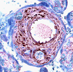
- Experimental details
- FGF-9 in Human Placenta. FGF-9 was detected in immersion fixed paraffin-embedded sections of human placenta using 5 µg/mL Goat Anti-Human FGF-9 Antigen Affinity-purified Polyclonal Antibody (Catalog # AF-273-NA) overnight at 4 °C. Tissue was stained with the Anti-Goat HRP-AEC Cell & Tissue Staining Kit (red; Catalog # CTS009) and counterstained with hematoxylin (blue). View our protocol for Chromogenic IHC Staining of Paraffin-embedded Tissue Sections.
