Explore
 Validate
 Learn
Learn Western blot
 Immunocytochemistry
Antibody data
- Antibody Data
- Antigen structure
- References [1]
- Comments [0]
- Validations
- Western blot [2]
- Chromatin Immunoprecipitation [1]
Submit
Validation data
Reference
Comment
Report error
- Product number
- AF2699 - Provider product page

- Provider
- Novus Biologicals
- Product name
- Goat Polyclonal c-Rel Antibody
- Antibody type
- Polyclonal
- Description
- Antigen Affinity-purified. Detects human and mouse c-Rel in Western blots.
- Reactivity
- Human, Mouse
- Host
- Goat
- Conjugate
- Unconjugated
- Isotype
- IgG
- Vial size
- 100 ug
- Concentration
- LYOPH
- Storage
- Use a manual defrost freezer and avoid repeated freeze-thaw cycles. 12 months from date of receipt, -20 to -70 degreesC as supplied. 1 month, 2 to 8 degreesC under sterile conditions after reconstitution. 6 months, -20 to -70 degreesC under sterile conditions after reconstitution.
Submitted references TNFR-associated factor 2 deficiency in B lymphocytes predisposes to chronic lymphocytic leukemia/small lymphocytic lymphoma in mice.
Pérez-Chacón G, Llobet D, Pardo C, Pindado J, Choi Y, Reed JC, Zapata JM
Journal of immunology (Baltimore, Md. : 1950) 2012 Jul 15;189(2):1053-61
Journal of immunology (Baltimore, Md. : 1950) 2012 Jul 15;189(2):1053-61
No comments: Submit comment
Supportive validation
- Submitted by
- Novus Biologicals (provider)
- Main image
- Experimental details
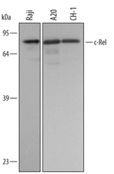
- Detection of Human/Mouse c-Rel by Western Blot. Western blot shows lysates of Raji human Burkitt's lymphoma cell line, A20 mouse B cell lymphoma cell line, and CH-1 mouse B cell lymphoma cell line. PVDF membrane was probed with 0.5 µg/mL of Goat Anti-Human/Mouse c-Rel Antigen Affinity-purified Polyclonal Antibody (Catalog # AF2699) followed by HRP-conjugated Anti-Goat IgG Secondary Antibody (Catalog # HAF109). A specific band was detected for c-Rel at approximately 69 kDa (as indicated). This experiment was conducted under reducing conditions and using Immunoblot Buffer Group 1.
- Submitted by
- Novus Biologicals (provider)
- Main image

- Experimental details
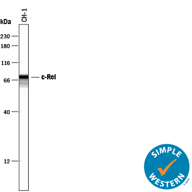
- Detection of Mouse c-Rel by Simple WesternTM. Simple Western lane view shows lysates of CH-1 mouse B cell lymphoma cell line, loaded at 0.2 mg/mL. A specific band was detected for c-Rel at approximately 71 kDa (as indicated) using 5 µg/mL of Goat Anti-Human/Mouse c-Rel Antigen Affinity-purified Polyclonal Antibody (Catalog # AF2699) followed by 1:50 dilution of HRP-conjugated Anti-Goat IgG Secondary Antibody (Catalog # HAF109). This experiment was conducted under reducing conditions and using the 12-230 kDa separation system.
Supportive validation
- Submitted by
- Novus Biologicals (provider)
- Main image
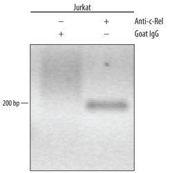
- Experimental details
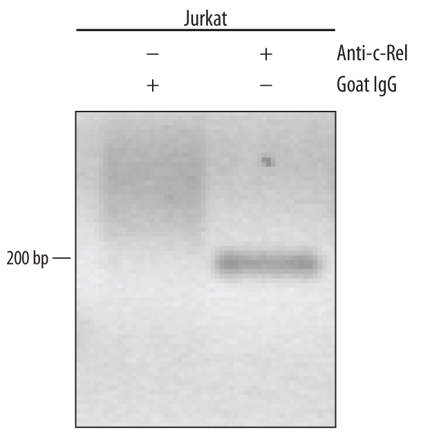
- Detection of c-Rel-regulated Genes by Chromatin Immunoprecipitation. Jurkat human acute T cell leukemia cell line treated with 50 ng/mL and 200 ng/mL PMA and calcium ionomycin for overnight was fixed using formaldehyde, resuspended in lysis buffer, and sonicated to shear chromatin. c-Rel/DNA complexes were immuno-precipitated using 5 μg Goat Anti-Human/Mouse c-Rel Antigen Affinity-purified Polyclonal Antibody (Catalog # AF2699) or control antibody (Catalog # AB-108-C) for 15 minutes in an ultrasonic bath, followed by Biotinylated Anti-Goat IgG Secondary Antibody (Catalog # BAF109). Immunocomplexes were captured using 50 μL of MagCellect Streptavidin Ferrofluid (Catalog # MAG999) and DNA was purified using chelating resin solution. The p21 promoter was detected by standard PCR.