 Explore
 Validate
 Learn
Learn Western blot
Antibody data
- Antibody Data
- Antigen structure
- References [0]
- Comments [0]
- Validations
- Western blot [3]
- Immunocytochemistry [1]
- Chromatin Immunoprecipitation [3]
Submit
Validation data
Reference
Comment
Report error
- Product number
- PA5-47370 - Provider product page

- Provider
- Invitrogen Antibodies
- Product name
- c-Rel Polyclonal Antibody
- Antibody type
- Polyclonal
- Antigen
- Recombinant full-length protein
- Description
- Reconstitute at 0.2 mg/mL in sterile PBS.
- Reactivity
- Human, Mouse
- Host
- Goat
- Isotype
- IgG
- Vial size
- 100 µg
- Concentration
- 0.2 mg/mL
- Storage
- -20° C, Avoid Freeze/Thaw Cycles
No comments: Submit comment
Supportive validation
- Submitted by
- Invitrogen Antibodies (provider)
- Main image

- Experimental details
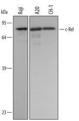
- Western blot analysis from lysates of Raji human Burkitt's lymphoma cell line, A20 mouse B cell lymphoma cell line, and CH-1 mouse B cell lymphoma cell line. PVDF membrane was probed with 0.5 µg/mL of Goat Anti-human/mouse c-Rel Antigen Affinity-purified Polyclonal Antibody (Product # PA5-47370) followed by HRP-conjugated Anti-Goat IgG Secondary Antibody. A specific band was detected for c-Rel at approximately 69 kDa (as indicated). This experiment was conducted under reducing conditions.

- Submitted by
- Invitrogen Antibodies (provider)
- Main image
- Experimental details
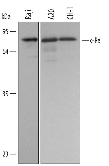
- Western blot analysis of c-Rel in Raji human Burkitts lymphoma cell line, A20 mouse B cell lymphoma cell line, and CH‚1 mouse B cell lymphoma cell line. Samples were incubated in c-Rel polyclonal antibody (Product # PA5-47370) using a dilution of 0.5 µg/mL followed by a HRP-conjugated Anti-Goat IgG secondary antibody. A specific band was detected for c-Rel at approximately 69 kDa (as indicated). This experiment was conducted under reducing conditions.
- Submitted by
- Invitrogen Antibodies (provider)
- Main image

- Experimental details
- Western blot analysis of c-Rel in 0.2 mg/mL lysates of CH‚1 mouse B cell lymphoma cell line. Samples were incubated in c-Rel polyclonal antibody (Product # PA5-47370) using a dilution of 5 µg/mL followed by HRP-conjugated Anti-Goat IgG at a dilution of 0.0763888888888889. A specific band was detected for c‚Rel at approximately 71 kDa (as indicated). This experiment was conducted under reducing conditions and using the 12-230 kDa separation system.

Supportive validation
- Submitted by
- Invitrogen Antibodies (provider)
- Main image

- Experimental details
- Immunocytochemistry analysis of c-Rel in immersion fixed THP‚1 human acute monocytic leukemia cell line treated with LPS. Samples were incubated in c-Rel polyclonal antibody (Product # PA5-47370) using a dilution of 5 µg/mL for 3 hours at room temperature followed by NorthernLights™ 557-conjugated Anti-Goat IgG Secondary Antibody (red) and counterstained with DAPI (blue). Specific staining was localized to cell nuclei.

Supportive validation
- Submitted by
- Invitrogen Antibodies (provider)
- Main image

- Experimental details
- Chromatin immunoprecipitation of Jurkat human acute T cell leukemia cell line treated with 50 ng/mL and 200 ng/mL PMA and calcium ionomycin for overnight was fixed using formaldehyde, resuspended in lysis buffer, and sonicated to shear chromatin. c-Rel/DNA complexes were immuno-precipitated using 5 µg Goat Anti-human/mouse c-Rel Antigen Affinity-purified Polyclonal Antibody (Product # PA5-47370) or control antibody for 15 minutes in an ultrasonic bath, followed by Biotinylated Anti-Goat IgG Secondary Antibody. Immunocomplexes were captured using 50 µL of MagCellect Streptavidin Ferrofluid and DNA was purified using chelating resin solution. The p21 promoter was detected by standard PCR.

- Submitted by
- Invitrogen Antibodies (provider)
- Main image

- Experimental details
- ChIP assay of c-Rel in Jurkat human acute T cell leukemia cell line. Samples were immunoprecipitated with c-Rel polyclonal antibody (Product # PA5-47370) for 15 minutes in an ultrasonic bath using a dilution of 5 μg followed by a Biotinylated Anti-Goat IgG secondary antibody. Cell line was treated with 50 ng/mL and 200 ng/mL PMA and calcium ionomycin for overnight was fixed using formaldehyde, resuspended in lysis buffer, and sonicated to shear chromatin. Immunocomplexes were captured using 50 μL of MagCellect Streptavidin Ferrofluid and DNA was purified using chelating resin solution. The p21 promoter was detected by standard PCR.
