 Explore
 Validate
 Learn
Learn Western blot
 Immunohistochemistry
Antibody data
- Antibody Data
- Antigen structure
- References [0]
- Comments [0]
- Validations
- Western blot [7]
- Immunohistochemistry [17]
Submit
Validation data
Reference
Comment
Report error
- Product number
- LS-C114449 - Provider product page

- Provider
- LSBio
- Product name
- GAD1 / GAD67 Antibody (clone OTI3G9) LS-C114449
- Antibody type
- Monoclonal
- Description
- Affinity purified
- Reactivity
- Human
- Host
- Mouse
- Isotype
- IgG
- Antibody clone number
- OTI3G9
- Storage
- Store at -20°C. Avoid freeze-thaw cycles.
No comments: Submit comment
Supportive validation
- Submitted by
- LSBio (provider)
- Enhanced method
- Genetic validation
- Main image

- Experimental details
- Western blot analysis of 35ug of cell extracts from canine Kidney (MDCK) cells using anti-GAD1 antibody.

- Submitted by
- LSBio (provider)
- Enhanced method
- Genetic validation
- Main image

- Experimental details
- Western blot analysis of 35ug of cell extracts from human Kidney (HEK293T) cells using anti-GAD1 antibody.

- Submitted by
- LSBio (provider)
- Enhanced method
- Genetic validation
- Main image

- Experimental details
- Western blot analysis of 35ug of cell extracts from Green monkey Kiney (COS7) cells using anti-GAD1 antibody.

- Submitted by
- LSBio (provider)
- Enhanced method
- Genetic validation
- Main image

- Experimental details
- Western blot analysis of 35ug of cell extracts from human Lung adenocarcinoma (A549) cells using anti-GAD1 antibody.

- Submitted by
- LSBio (provider)
- Enhanced method
- Genetic validation
- Main image

- Experimental details
- Western blot analysis of 35ug of cell extracts from human colon adenocarcinoma (HT29) cells using anti-GAD1 antibody.

- Submitted by
- LSBio (provider)
- Enhanced method
- Genetic validation
- Main image

- Experimental details
- Western blot analysis of 35ug of cell extracts from human (HeLa) cells using anti-GAD1 antibody.

- Submitted by
- LSBio (provider)
- Enhanced method
- Genetic validation
- Main image

- Experimental details
- HEK293T cells were transfected with the pCMV6-ENTRY control (Left lane) or pCMV6-ENTRY GAD1 (Right lane) cDNA for 48 hrs and lysed. Equivalent amounts of cell lysates (5 ug per lane) were separated by SDS-PAGE and immunoblotted with anti-GAD1.

Enhanced validation
- Submitted by
- LSBio (provider)
- Enhanced method
- Genetic validation
- Main image

- Experimental details
- Immunohistochemical staining of paraffin-embedded colon tissue using anti-GAD1 mouse monoclonal antibody. (Dilution 1:50).

- Submitted by
- LSBio (provider)
- Enhanced method
- Genetic validation
- Main image
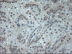
- Experimental details
- Immunohistochemical staining of paraffin-embedded Carcinoma of kidney tissue using anti-GAD1 mouse monoclonal antibody. (Dilution 1:50).

- Submitted by
- LSBio (provider)
- Enhanced method
- Genetic validation
- Main image

- Experimental details
- Immunohistochemical staining of paraffin-embedded lung tissue using anti-GAD1 mouse monoclonal antibody. (Dilution 1:50).

- Submitted by
- LSBio (provider)
- Enhanced method
- Genetic validation
- Main image

- Experimental details
- Immunohistochemical staining of paraffin-embedded Ovary tissue using anti-GAD1 mouse monoclonal antibody. (Dilution 1:50).

- Submitted by
- LSBio (provider)
- Enhanced method
- Genetic validation
- Main image

- Experimental details
- Immunohistochemical staining of paraffin-embedded Adenocarcinoma of ovary tissue using anti-GAD1 mouse monoclonal antibody. (Dilution 1:50).

- Submitted by
- LSBio (provider)
- Enhanced method
- Genetic validation
- Main image

- Experimental details
- Immunohistochemical staining of paraffin-embedded pancreas tissue using anti-GAD1 mouse monoclonal antibody. (Dilution 1:50).

- Submitted by
- LSBio (provider)
- Enhanced method
- Genetic validation
- Main image

- Experimental details
- Immunohistochemical staining of paraffin-embedded thyroid tissue using anti-GAD1 mouse monoclonal antibody. (Dilution 1:50).

- Submitted by
- LSBio (provider)
- Enhanced method
- Genetic validation
- Main image

- Experimental details
- Immunohistochemical staining of paraffin-embedded pancreas tissue using anti-GAD1 mouse monoclonal antibody. (Dilution 1:50).

- Submitted by
- LSBio (provider)
- Enhanced method
- Genetic validation
- Main image

- Experimental details
- Immunohistochemical staining of paraffin-embedded Adenocarcinoma of ovary tissue using anti-GAD1 mouse monoclonal antibody. (Dilution 1:50).

- Submitted by
- LSBio (provider)
- Enhanced method
- Genetic validation
- Main image

- Experimental details
- Immunohistochemical staining of paraffin-embedded Ovary tissue using anti-GAD1 mouse monoclonal antibody. (Dilution 1:50).

- Submitted by
- LSBio (provider)
- Enhanced method
- Genetic validation
- Main image

- Experimental details
- Immunohistochemical staining of paraffin-embedded lung tissue using anti-GAD1 mouse monoclonal antibody. (Dilution 1:50).

- Submitted by
- LSBio (provider)
- Enhanced method
- Genetic validation
- Main image

- Experimental details
- Immunohistochemical staining of paraffin-embedded endometrium tissue using anti-GAD1 mouse monoclonal antibody. (Dilution 1:50).

- Submitted by
- LSBio (provider)
- Enhanced method
- Genetic validation
- Main image

- Experimental details
- Immunohistochemical staining of paraffin-embedded Carcinoma of bladder tissue using anti-GAD1 mouse monoclonal antibody. (Dilution 1:50).

- Submitted by
- LSBio (provider)
- Enhanced method
- Genetic validation
- Main image

- Experimental details
- Immunohistochemical staining of paraffin-embedded Adenocarcinoma of breast tissue using anti-GAD1 mouse monoclonal antibody. (Dilution 1:50).

- Submitted by
- LSBio (provider)
- Main image
- Experimental details
- Immunohistochemical staining of paraffin-embedded Adenocarcinoma of breast tissue using anti-GAD1 mouse monoclonal antibody. (Dilution 1:50).

- Submitted by
- LSBio (provider)
- Main image

- Experimental details
- Immunohistochemical staining of paraffin-embedded colon tissue using anti-GAD1 mouse monoclonal antibody. (Dilution 1:50).

- Submitted by
- LSBio (provider)
- Main image

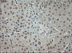
- Experimental details
- Immunohistochemical staining of paraffin-embedded Carcinoma of kidney tissue using anti-GAD1 mouse monoclonal antibody. (Dilution 1:50).
