Explore
 Validate
 Learn
Learn Western blot
 Immunocytochemistry
Antibody data
- Antibody Data
- Antigen structure
- References [3]
- Comments [0]
- Validations
- Immunocytochemistry [4]
- Immunohistochemistry [8]
- Other assay [3]
Submit
Validation data
Reference
Comment
Report error
- Product number
- PA5-21397 - Provider product page

- Provider
- Invitrogen Antibodies
- Product name
- GAD67 Polyclonal Antibody
- Antibody type
- Polyclonal
- Antigen
- Recombinant full-length protein
- Description
- Recommended positive controls: SK-N-SH, mouse brain, rat brain, human GAD1-transfected 293T. Predicted reactivity: Mouse (96%), Rat (96%), Dog (99%), Cat (99%), Pig (99%), Chicken (90%), Chimpanzee (100%), Bovine (98%). Store product as a concentrated solution. Centrifuge briefly prior to opening the vial.
- Reactivity
- Human, Mouse, Rat
- Host
- Rabbit
- Isotype
- IgG
- Vial size
- 100 μL
- Concentration
- 0.22 mg/mL
- Storage
- Store at 4°C short term. For long term storage, store at -20°C, avoiding freeze/thaw cycles.
Submitted references Screening Biophysical Sensors and Neurite Outgrowth Actuators in Human Induced-Pluripotent-Stem-Cell-Derived Neurons.
Overexpression of Glutamate Decarboxylase in Mesenchymal Stem Cells Enhances Their Immunosuppressive Properties and Increases GABA and Nitric Oxide Levels.
Region-specific effects of repeated ketamine administration on the presynaptic GABAergic neurochemistry in rat brain.
Pai VP, Cooper BG, Levin M
Cells 2022 Aug 9;11(16)
Cells 2022 Aug 9;11(16)
Overexpression of Glutamate Decarboxylase in Mesenchymal Stem Cells Enhances Their Immunosuppressive Properties and Increases GABA and Nitric Oxide Levels.
Urrutia M, Fernández S, González M, Vilches R, Rojas P, Vásquez M, Kurte M, Vega-Letter AM, Carrión F, Figueroa F, Rojas P, Irarrázabal C, Fuentealba RA
PloS one 2016;11(9):e0163735
PloS one 2016;11(9):e0163735
Region-specific effects of repeated ketamine administration on the presynaptic GABAergic neurochemistry in rat brain.
Boczek T, Lisek M, Ferenc B, Wiktorska M, Ivchevska I, Zylinska L
Neurochemistry international 2015 Dec;91:13-25
Neurochemistry international 2015 Dec;91:13-25
No comments: Submit comment
Supportive validation
- Submitted by
- Invitrogen Antibodies (provider)
- Main image
- Experimental details
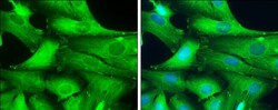
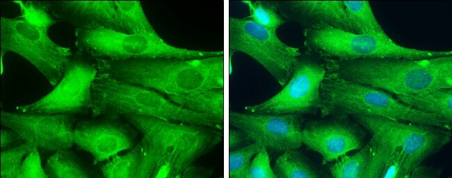
- Immunocytochemistry-Immunofluorescence analysis of GAD67 was performed in SK-N-SH cells fixed in 4% paraformaldehyde at RT for 15 min. Green: GAD67 Polyclonal Antibody (Product # PA5-21397) diluted at 1:200. Blue: Hoechst 33342 staining.
- Submitted by
- Invitrogen Antibodies (provider)
- Main image

- Experimental details
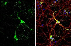
- GAD67 Polyclonal Antibody detects GAD67 protein by immunofluorescent analysis. Sample: DIV10 rat E18 primary cortical neuron cells were fixed in 4% paraformaldehyde at RT for 15 min. Green: GAD67 stained by GAD67 Polyclonal Antibody (Product # PA5-21397) diluted at 1:500. Red: Tau, stained by Phospho-Tau (Ser262) Polyclonal Antibody [GT287]diluted at 1:500. Blue: Fluoroshield with DAPI .

- Submitted by
- Invitrogen Antibodies (provider)
- Main image
- Experimental details
- GAD67 Polyclonal Antibody detects GAD67 protein by immunofluorescent analysis. Sample: DIV10 rat E18 primary cortical neuron cells were fixed in 4% paraformaldehyde at RT for 15 min. Green: GAD67 stained by GAD67 Polyclonal Antibody (Product # PA5-21397) diluted at 1:500. Red: Tau, stained by Phospho-Tau (Ser262) Polyclonal Antibody [GT287]diluted at 1:500. Blue: Fluoroshield with DAPI .

- Submitted by
- Invitrogen Antibodies (provider)
- Main image

- Experimental details
- Immunocytochemistry-Immunofluorescence analysis of GAD67 was performed in SK-N-SH cells fixed in 4% paraformaldehyde at RT for 15 min. Green: GAD67 Polyclonal Antibody (Product # PA5-21397) diluted at 1:200. Blue: Hoechst 33342 staining.

Supportive validation
- Submitted by
- Invitrogen Antibodies (provider)
- Main image
- Experimental details
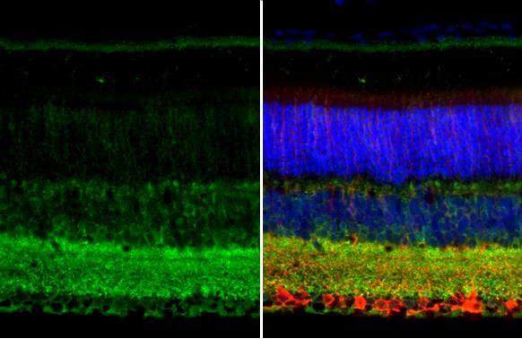
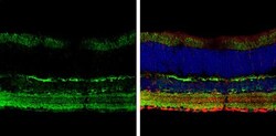
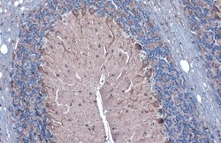
- GAD67 Polyclonal Antibody detects GAD67 protein at cell membrane and cytoplasm by immunohistochemical analysis. Sample: Paraffin-embedded mouse eye. Green: GAD67 stained by GAD67 Polyclonal Antibody (Product # PA5-21397) diluted at 1:250. Red: beta Tubulin 3/ Tuj1, a neural marker, stained by beta Tubulin 3/ Tuj1 antibody [GT11710] diluted at 1:500. Blue: Fluoroshield with DAPI. Antigen Retrieval: Citrate buffer, pH 6.0, 15 min.
- Submitted by
- Invitrogen Antibodies (provider)
- Main image
- Experimental details
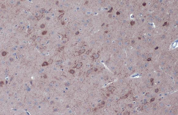
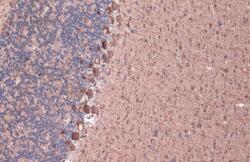
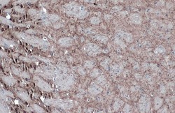
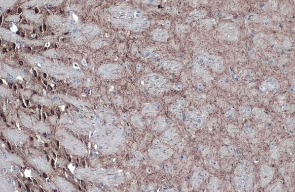
- GAD67 Polyclonal Antibody detects GAD67 protein at cytoplasm by immunohistochemical analysis. Sample: Paraffin-embedded rat brain. GAD67 stained by GAD67 Polyclonal Antibody (Product # PA5-21397) diluted at 1:500. Antigen Retrieval: Citrate buffer, pH 6.0, 15 min.
- Submitted by
- Invitrogen Antibodies (provider)
- Main image
- Experimental details
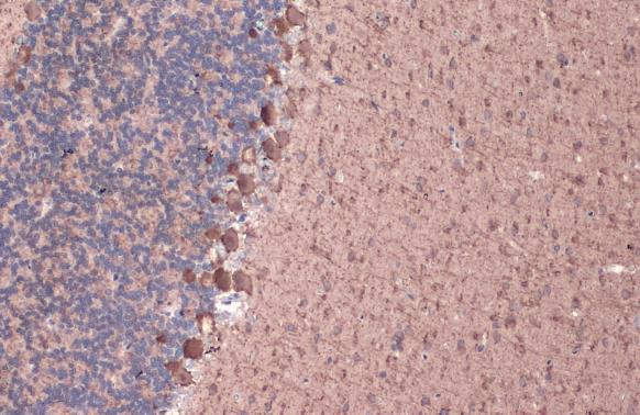
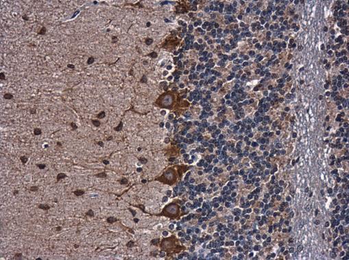
- GAD67 Polyclonal Antibody detects GAD67 protein at cell membrane and cytoplasm by immunohistochemical analysis. Sample: Paraffin-embedded rat cerebellum. GAD67 stained by GAD67 Polyclonal Antibody (Product # PA5-21397) diluted at 1:1,000. Antigen Retrieval: Citrate buffer, pH 6.0, 15 min.
- Submitted by
- Invitrogen Antibodies (provider)
- Main image
- Experimental details
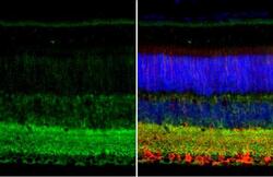
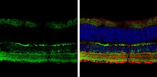
- Immunohistochemistry (Frozen) analysis of GAD67 was performed in frozen sectioned adult mouse retina tissue using GAD67 Polyclonal Antibody (Product # PA5-21397) at a dilution of 1:250 (Green). Red: beta Tubulin 3/ TUJ1, stained by beta Tubulin 3/ TUJ1 antibody diluted at 1:250. Blue: Fluoroshield with DAPI.
- Submitted by
- Invitrogen Antibodies (provider)
- Main image

- Experimental details
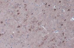
- Immunohistochemistry (Paraffin) analysis of GAD67 was performed in paraffin-embedded rat brain tissue using GAD67 Polyclonal Antibody (Product # PA5-21397) at a dilution of 1:500.
- Submitted by
- Invitrogen Antibodies (provider)
- Main image
- Experimental details
- GAD67 Polyclonal Antibody detects GAD67 protein at cytoplasm by immunohistochemical analysis. Sample: Paraffin-embedded mouse brain. GAD67 stained by GAD67 Polyclonal Antibody (Product # PA5-21397) diluted at 1:500. Antigen Retrieval: Citrate buffer, pH 6.0, 15 min.
- Submitted by
- Invitrogen Antibodies (provider)
- Main image
- Experimental details
- GAD67 Polyclonal Antibody detects GAD67 protein at cell membrane and cytoplasm by immunohistochemical analysis. Sample: Paraffin-embedded mouse cerebellum. GAD67 stained by GAD67 Polyclonal Antibody (Product # PA5-21397) diluted at 1:1,000. Antigen Retrieval: Citrate buffer, pH 6.0, 15 min.

- Submitted by
- Invitrogen Antibodies (provider)
- Main image

- Experimental details
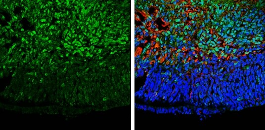
- Immunohistochemistry (Frozen) analysis of GAD67 was performed in frozen sectioned E13.5 Rat brain tissue using GAD67 Polyclonal Antibody (Product # PA5-21397) at a dilution of 1:250 (Green). Red: beta Tubulin 3/ TUJ1, a mature neuron marker, stained by beta Tubulin 3/ TUJ1 antibody diluted at 1:500. Blue: Fluoroshield with DAPI.
Supportive validation
- Submitted by
- Invitrogen Antibodies (provider)
- Main image
- Experimental details
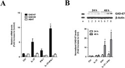
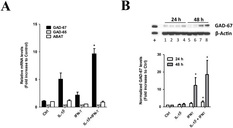
- Fig 3 GAD-67 gene expression regulation by pro-inflammatory cytokines IL-1beta and IFN-gamma in MSCs. MSC were seeded at 4,800 cells/cm 2 and grew for 24 h. MSC cultures were then supplemented with 20 ng/mL IL-1beta, 25 ng/mL IFN-gamma, or the combination of both cytokines, and MSC cells were grew for indicated times. Control cultures were identically treated but cytokines were not added. (A) GAD-67, GAD-65 and ABAT mRNA levels were determined 48 h after cytokine treatment by RT-qPCR. IL-1beta selectively increases GAD-67 mRNA levels in MSCs and IFN-gamma potentiates IL-1beta effects. *, p
- Submitted by
- Invitrogen Antibodies (provider)
- Main image
- Experimental details
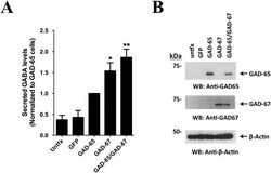
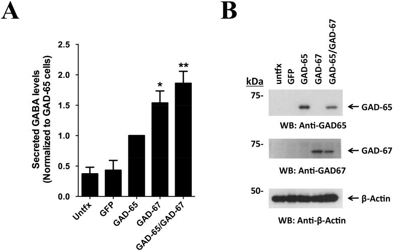
- Fig 4 Increased GABA secretion with GAD-65/GAD-67 co-expression. MSCs were seeded at 10,000 cells/cm 2 in 6 well plates and grew overnight. Cells were then transfected with Fugene6 as described in Methods using 1 mug total plasmid, and cells were recovered for 24 h. Co-expression with GAD-65 and GAD-67 used equivalent amounts of each plasmid. (A) Conditioned media was pre-cleared by centrifugation and secreted GABA levels were enzymatically determined using a fluorescence-coupled assay. Basal levels were 2.48 +- 0.83 muM and results were expressed as fold change to GAD-65 transfected (GAD-65) cells. Maximal secretion of GABA was detected using GADs co-expression. *, p
- Submitted by
- Invitrogen Antibodies (provider)
- Main image
- Experimental details
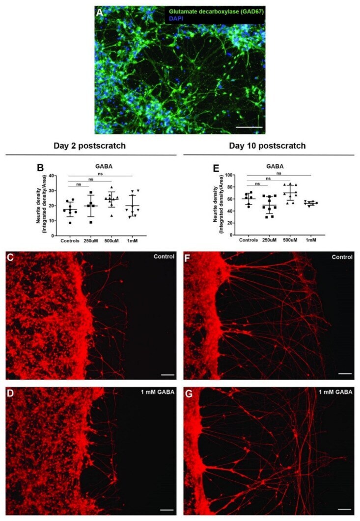
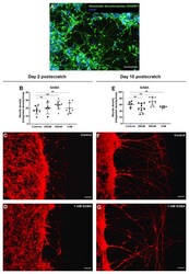
- Figure 7 GABA does not affect neurite outgrowth in scratch assay. ( A ) hiNSC-derived day 10 neuronal culture shows presence of GABAergic neurons (glutamate decarboxylase--GAD67 marker). ( B ) GABA treatment (48 h) shows no significant change in scratch neurite density on day 2 post-scratch. ( C , D ) Representative images of Calcein Red-Orange AM stained control neural cultures ( C ) and neural cultures treated with 1 mM GABA ( D ) on day 2 post-scratch showing no discernable change in neurite outgrowths with GABA treatment. ( E ) GABA treatment (48 h) shows no significant change in scratch neurite density on day 10 post-scratch. ( F , G ) Representative images of Calcein Red-Orange AM stained control neural cultures ( F ) and neural cultures treated with 1 mM GABA ( G ) on day 10 post-scratch showing no discernable change in neurite outgrowth with GABA treatment. All data are represented as mean +- S.D. ns--not significant. All scale bars, 100 um.