Explore
 Validate
 Learn
Learn Western blot
Antibody data
- Antibody Data
- Antigen structure
- References [4]
- Comments [0]
- Validations
- Western blot [1]
- Immunocytochemistry [1]
- Flow cytometry [1]
Submit
Validation data
Reference
Comment
Report error
- Product number
- AF2157 - Provider product page

- Provider
- R&D Systems
- Product name
- Human/Mouse/Rat GSK-3 alpha/beta Antibody
- Antibody type
- Polyclonal
- Description
- Immunogen affinity purified. Detects human, mouse, and rat GSK-3 beta and the closely related GSK-3 alpha .
- Reactivity
- Human, Mouse, Rat
- Host
- Rabbit
- Conjugate
- Unconjugated
- Antigen sequence
P49841- Isotype
- IgG
- Vial size
- 50 ug
- Concentration
- LYOPH
- Storage
- Use a manual defrost freezer and avoid repeated freeze-thaw cycles. 12 months from date of receipt, -20 to -70 °C as supplied. 1 month, 2 to 8 °C under sterile conditions after reconstitution. 6 months, -20 to -70 °C under sterile conditions after reconstitution.
Submitted references A ZIP6-ZIP10 heteromer controls NCAM1 phosphorylation and integration into focal adhesion complexes during epithelial-to-mesenchymal transition.
Mycobacteria bypass mucosal NF-kB signalling to induce an epithelial anti-inflammatory IL-22 and IL-10 response.
GSK3β regulates physiological migration of stem/progenitor cells via cytoskeletal rearrangement.
Survival effect of PDGF-CC rescues neurons from apoptosis in both brain and retina by regulating GSK3beta phosphorylation.
Brethour D, Mehrabian M, Williams D, Wang X, Ghodrati F, Ehsani S, Rubie EA, Woodgett JR, Sevalle J, Xi Z, Rogaeva E, Schmitt-Ulms G
Scientific reports 2017 Jan 18;7:40313
Scientific reports 2017 Jan 18;7:40313
Mycobacteria bypass mucosal NF-kB signalling to induce an epithelial anti-inflammatory IL-22 and IL-10 response.
Lutay N, Håkansson G, Alaridah N, Hallgren O, Westergren-Thorsson G, Godaly G
PloS one 2014;9(1):e86466
PloS one 2014;9(1):e86466
GSK3β regulates physiological migration of stem/progenitor cells via cytoskeletal rearrangement.
Lapid K, Itkin T, D'Uva G, Ovadya Y, Ludin A, Caglio G, Kalinkovich A, Golan K, Porat Z, Zollo M, Lapidot T
The Journal of clinical investigation 2013 Apr;123(4):1705-17
The Journal of clinical investigation 2013 Apr;123(4):1705-17
Survival effect of PDGF-CC rescues neurons from apoptosis in both brain and retina by regulating GSK3beta phosphorylation.
Tang Z, Arjunan P, Lee C, Li Y, Kumar A, Hou X, Wang B, Wardega P, Zhang F, Dong L, Zhang Y, Zhang SZ, Ding H, Fariss RN, Becker KG, Lennartsson J, Nagai N, Cao Y, Li X
The Journal of experimental medicine 2010 Apr 12;207(4):867-80
The Journal of experimental medicine 2010 Apr 12;207(4):867-80
No comments: Submit comment
Supportive validation
- Submitted by
- R&D Systems (provider)
- Main image
- Experimental details
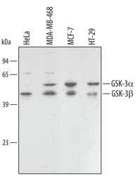
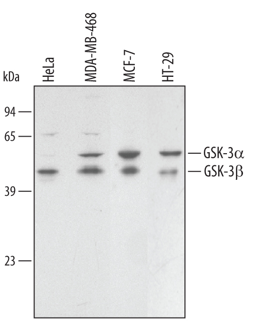
- Detection of Human GSK-3 alpha/beta by Western Blot. Western blot shows lysates of HeLa human cervical epithelial carcinoma cell line, HT-29 human colon adenocarcinoma cell line, MDA-MB-468 and MCF-7 human breast cancer cell line. PVDF membrane was probed with 0.1 µg/mL of Rabbit Anti-Human/Mouse/ Rat GSK-3 alpha/beta Antigen Affinity-purified Polyclonal Antibody (Catalog # AF2157) followed by HRP-conjugated Anti-Rabbit IgG Secondary Antibody (Catalog # HAF008). Specific bands were detected for GSK-3 alpha/beta at approximately 46 and 51 kDa (as indicated). This experiment was conducted under reducing conditions and using Immunoblot Buffer Group 1.
Supportive validation
- Submitted by
- R&D Systems (provider)
- Main image
- Experimental details
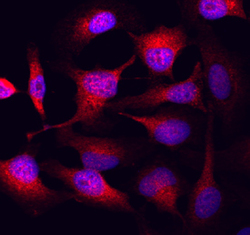
- GSK-3 alpha/beta in HeLa Human Cell Line. GSK-3 alpha/beta was detected in immersion fixed HeLa human cervical epithelial carcinoma cell line using Rabbit Anti-Human/Mouse/Rat GSK-3 alpha/beta Antigen Affinity-purified Polyclonal Antibody (Catalog # AF2157) at 10 µg/mL for 3 hours at room temperature. Cells were stained using the NorthernLights™ 557-conjugated Anti-Rabbit IgG Secondary Antibody (red; Catalog # NL004) and counterstained with DAPI (blue). Specific staining was localized to cytoplasm. View our protocol for Fluorescent ICC Staining of Cells on Coverslips.

Supportive validation
- Submitted by
- R&D Systems (provider)
- Main image
- Experimental details
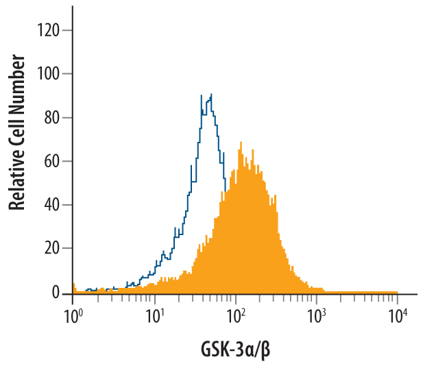
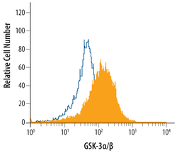
- Detection of GSK-3 alpha/beta in HeLa Human Cell Line by Flow Cytometry. HeLa human cervical epithelial carcinoma cell line was stained with Rabbit Anti-Human/Mouse/Rat GSK-3 alpha/beta Antigen Affinity-purified Polyclonal Antibody (Catalog # AF2157, filled histogram) or control antibody (Catalog # AB-105-C, open histogram), followed by Allophycocyanin-conjugated Anti-Rabbit IgG Secondary Antibody (Catalog # F0111). To facilitate intracellular staining, cells were fixed with paraformaldehyde and permeabilized with saponin.