Explore
 Validate
 Learn
Learn Western blot
 Immunocytochemistry
Antibody data
- Antibody Data
- Antigen structure
- References [4]
- Comments [0]
- Validations
- Western blot [1]
- Flow cytometry [1]
Submit
Validation data
Reference
Comment
Report error
- Product number
- AF1590 - Provider product page

- Provider
- R&D Systems
- Product name
- Human/Mouse/Rat Phospho-GSK-3 alpha/beta (S21/S9) Antibody
- Antibody type
- Polyclonal
- Description
- Antigen and protein A Affinity-purified. Detects human, mouse, and rat GSK-3 beta when phosphorylated at S9, and human, mouse, and rat GSK-3 alpha when phosphorylated at S21.
- Reactivity
- Human, Mouse, Rat
- Host
- Rabbit
- Conjugate
- Unconjugated
- Isotype
- IgG
- Vial size
- 100 ug
- Concentration
- LYOPH
- Storage
- Use a manual defrost freezer and avoid repeated freeze-thaw cycles. 12 months from date of receipt, -20 to -70 °C as supplied. 1 month, 2 to 8 °C under sterile conditions after reconstitution. 6 months, -20 to -70 °C under sterile conditions after reconstitution.
Submitted references HYS-32-Induced Microtubule Catastrophes in Rat Astrocytes Involves the PI3K-GSK3beta Signaling Pathway.
Mycobacteria bypass mucosal NF-kB signalling to induce an epithelial anti-inflammatory IL-22 and IL-10 response.
Mechanical induction of PGE2 in osteocytes blocks glucocorticoid-induced apoptosis through both the β-catenin and PKA pathways.
Survival effect of PDGF-CC rescues neurons from apoptosis in both brain and retina by regulating GSK3beta phosphorylation.
Chiu CT, Liao CK, Shen CC, Tang TK, Jow GM, Wang HS, Wu JC
PloS one 2015;10(5):e0126217
PloS one 2015;10(5):e0126217
Mycobacteria bypass mucosal NF-kB signalling to induce an epithelial anti-inflammatory IL-22 and IL-10 response.
Lutay N, Håkansson G, Alaridah N, Hallgren O, Westergren-Thorsson G, Godaly G
PloS one 2014;9(1):e86466
PloS one 2014;9(1):e86466
Mechanical induction of PGE2 in osteocytes blocks glucocorticoid-induced apoptosis through both the β-catenin and PKA pathways.
Kitase Y, Barragan L, Qing H, Kondoh S, Jiang JX, Johnson ML, Bonewald LF
Journal of bone and mineral research : the official journal of the American Society for Bone and Mineral Research 2010 Dec;25(12):2657-68
Journal of bone and mineral research : the official journal of the American Society for Bone and Mineral Research 2010 Dec;25(12):2657-68
Survival effect of PDGF-CC rescues neurons from apoptosis in both brain and retina by regulating GSK3beta phosphorylation.
Tang Z, Arjunan P, Lee C, Li Y, Kumar A, Hou X, Wang B, Wardega P, Zhang F, Dong L, Zhang Y, Zhang SZ, Ding H, Fariss RN, Becker KG, Lennartsson J, Nagai N, Cao Y, Li X
The Journal of experimental medicine 2010 Apr 12;207(4):867-80
The Journal of experimental medicine 2010 Apr 12;207(4):867-80
No comments: Submit comment
Supportive validation
- Submitted by
- R&D Systems (provider)
- Main image
- Experimental details
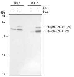
- Detection of Human Phospho-GSK-3 alpha (S21) and GSK-3 beta (S9) by Western Blot. Western blot shows lysates of HeLa human cervical epithelial carcinoma cell line untreated (-) or treated (+) with 200 nM PMA for 20 minutes and MCF-7 human breast cancer cell line untreated or treated with 100 ng/mL Recombinant Human IGF-1 (Catalog # 291-G1) for 15 minutes. PVDF membrane was probed with 0.2 µg/mL of Human/Mouse/Rat Phospho-GSK-3 alpha/beta (S21/S9) Antigen Affinity-purified Polyclonal Antibody (Catalog # AF1590), followed by HRP-conjugated Anti-Rabbit IgG Secondary Antibody (Catalog # HAF008). Specific bands were detected for Phospho-GSK-3 alpha (S21) and GSK-3 beta (S9) at approximately 51 and 46 kDa (as indicated). This experiment was conducted under reducing conditions and using Immunoblot Buffer Group 1.

Supportive validation
- Submitted by
- R&D Systems (provider)
- Main image
- Experimental details
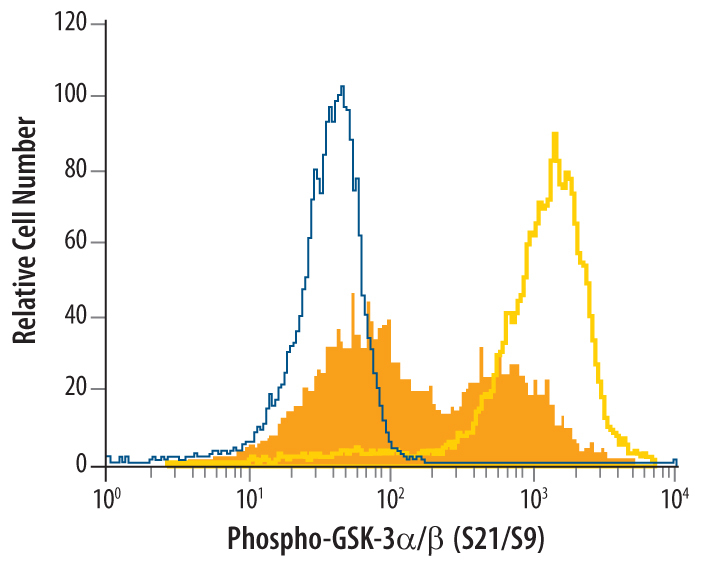
- Detection of Phospho-GSK-3 alpha/beta (S21/S9) in Jurkat Human Cell Line by Flow Cytometry. Jurkat human acute T cell leukemia cells, untreated (open histogram, yellow line), or treated with PI3-kinase inhibitors (dark orange filled histogram), were stained with Human/Mouse/Rat Phospho-GSK-3 alpha/beta (S21/S9) Antigen Affinity-purified Polyclonal Antibody (Catalog # AF1590) or control antibody (Catalog # AB-105-C, blue open histogram), followed by Phycoerythrin-conjugated Anti-Rabbit IgG Secondary Antibody (Catalog # F0110). To facilitate intracellular staining, cells were fixed with paraformaldehyde and permeabilized with saponin.