Explore
 Validate
 Learn
Learn Western blot
Antibody data
- Antibody Data
- Antigen structure
- References [2]
- Comments [0]
- Validations
- Western blot [6]
- Immunocytochemistry [2]
- Immunoprecipitation [1]
- Immunohistochemistry [2]
Submit
Validation data
Reference
Comment
Report error
- Product number
- GTX103278 - Provider product page

- Provider
- GeneTex
- Proper citation
- GeneTex Cat#GTX103278, RRID:AB_1950505
- Product name
- Homer1 antibody
- Antibody type
- Polyclonal
- Reactivity
- Human, Mouse, Rat
- Host
- Rabbit
Submitted references Homer2 within the nucleus accumbens core bidirectionally regulates alcohol intake by both P and Wistar rats.
Binge alcohol drinking by mice requires intact group 1 metabotropic glutamate receptor signaling within the central nucleus of the amygdala.
Haider A, Woodward NC, Lominac KD, Sacramento AD, Klugmann M, Bell RL, Szumlinski KK
Alcohol (Fayetteville, N.Y.) 2015 Sep;49(6):533-42
Alcohol (Fayetteville, N.Y.) 2015 Sep;49(6):533-42
Binge alcohol drinking by mice requires intact group 1 metabotropic glutamate receptor signaling within the central nucleus of the amygdala.
Cozzoli DK, Courson J, Wroten MG, Greentree DI, Lum EN, Campbell RR, Thompson AB, Maliniak D, Worley PF, Jonquieres G, Klugmann M, Finn DA, Szumlinski KK
Neuropsychopharmacology : official publication of the American College of Neuropsychopharmacology 2014 Jan;39(2):435-44
Neuropsychopharmacology : official publication of the American College of Neuropsychopharmacology 2014 Jan;39(2):435-44
No comments: Submit comment
Supportive validation
- Submitted by
- GeneTex (provider)
- Main image
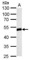
- Experimental details
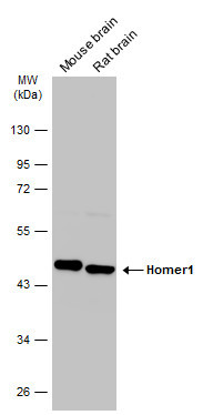
- Homer antibody detects HOMER1 protein by western blot analysis.A. 50 ?g rat brain lysate/extract 10% SDS-PAGEHomer antibody (GTX103278) dilution: 1:500 The HRP-conjugated anti-rabbit IgG antibody (GTX213110-01) was used to detect the primary antibody.

- Submitted by
- GeneTex (provider)
- Main image
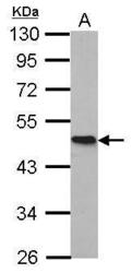
- Experimental details
- Homer antibody detects HOMER1 protein by western blot analysis.A. 30 ?g IMR32 whole cell lysate/extract10% SDS-PAGEHomer antibody (GTX103278) dilution: 1:1000 The HRP-conjugated anti-rabbit IgG antibody (GTX213110-01) was used to detect the primary antibody.

- Submitted by
- GeneTex (provider)
- Main image
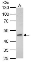
- Experimental details
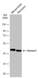
- Sample (20 ?g of whole cell lysate) A: mouse brain 10% SDS PAGE GTX103278 diluted at 1:20000 The HRP-conjugated anti-rabbit IgG antibody (GTX213110-01) was used to detect the primary antibody.

- Submitted by
- GeneTex (provider)
- Main image
- Experimental details
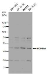
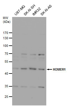
- Homer antibody detects Homer protein by western blot analysis. Various whole cell extracts (30 ?g) were separated by 10% SDS-PAGE, and blotted with Homer antibody (GTX103278) diluted by 1:1000. The HRP-conjugated anti-rabbit IgG antibody (GTX213110-01) was used to detect the primary antibody.
- Submitted by
- GeneTex (provider)
- Main image
- Experimental details
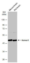
- Various tissue extracts (50 ?g) were separated by 10% SDS-PAGE, and the membrane was blotted with Homer1 antibody (GTX103278) diluted at 1:4000. The HRP-conjugated anti-rabbit IgG antibody (GTX213110-01) was used to detect the primary antibody.
- Submitted by
- GeneTex (provider)
- Main image
- Experimental details
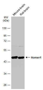
- Various tissue extracts (50 ?g) were separated by 10% SDS-PAGE, and the membrane was blotted with Homer1 antibody (GTX103278) diluted at 1:5000. The HRP-conjugated anti-rabbit IgG antibody (GTX213110-01) was used to detect the primary antibody.
Supportive validation
- Submitted by
- GeneTex (provider)
- Main image
- Experimental details
- Immunofluorescence analysis of methanol-fixed HeLa, using Homer(GTX103278) antibody at 1:500 dilution.

- Submitted by
- GeneTex (provider)
- Main image
- Experimental details
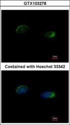
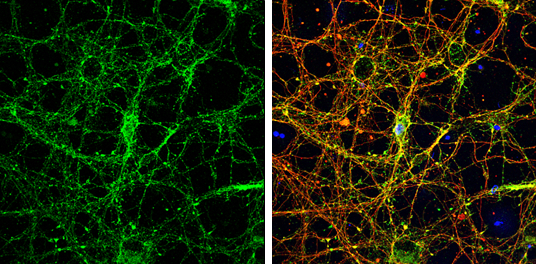
- Homer1 antibody detects Homer1 protein by immunofluorescent analysis.Sample: DIV9 rat E18 primary cortical neurons were fixed in 4% paraformaldehyde at RT for 15 min.Green: Homer1 protein stained by Homer1 antibody (GTX103278) diluted at 1:500.Red: beta Tubulin 3/ Tuj1, stained by beta Tubulin 3/ Tuj1 antibody [GT886] (GTX631830) diluted at 1:500.Blue: Fluoroshield with DAPI (GTX30920).
Supportive validation
- Submitted by
- GeneTex (provider)
- Main image
- Experimental details
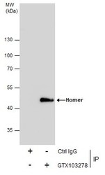
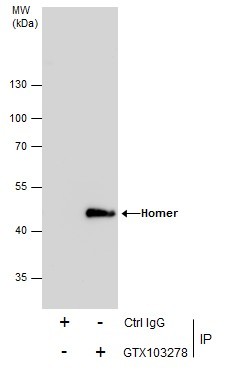
- Immunoprecipitation of Homer protein from IMR32 whole cell extracts using 5 £gg of Homer antibody (GTX103278).Western blot analysis was performed using Homer antibody (GTX103278).EasyBlot anti-Rabbit IgG (GTX221666-01) was used as a secondary reagent.
Supportive validation
- Submitted by
- GeneTex (provider)
- Main image
- Experimental details
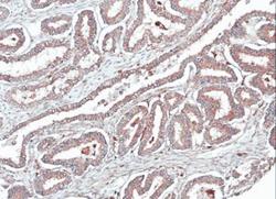
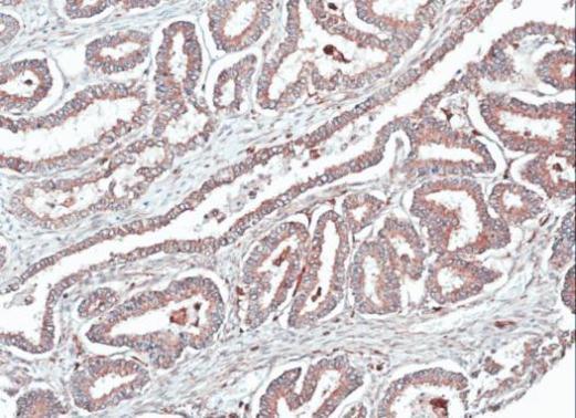
- Immunohistochemical analysis of paraffin-embedded human ovarian cancer, using HOMER1(GTX103278) antibody at 1:100 dilution.
- Submitted by
- GeneTex (provider)
- Main image
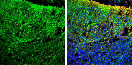
- Experimental details
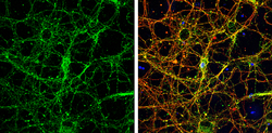
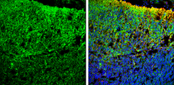
- Homer antibody detects Homer protein expression by immunohistochemical analysis.Sample: Frozen sectioned E13.5 Rat brain. Green: Homer protein stained by Homer antibody (GTX103278) diluted at 1:250.Red: beta Tubulin 3/ TUJ1, a mature neuron marker, stained by beta Tubulin 3/ TUJ1 antibody [GT11710] (GTX631836) diluted at 1:500.Blue: Fluoroshield with DAPI (GTX30920).