Explore
 Validate
 Learn
Learn Western blot
 Immunocytochemistry
Antibody data
- Antibody Data
- Antigen structure
- References [1]
- Comments [0]
- Validations
- Immunocytochemistry [7]
- Immunoprecipitation [1]
- Immunohistochemistry [2]
- Other assay [1]
Submit
Validation data
Reference
Comment
Report error
- Product number
- PA5-21487 - Provider product page

- Provider
- Invitrogen Antibodies
- Product name
- HOMER1 Polyclonal Antibody
- Antibody type
- Polyclonal
- Antigen
- Recombinant full-length protein
- Description
- Recommended positive controls: U87-MG, SK-N-SH, IMR32, SK-N-AS, mouse brain, rat brain. Predicted reactivity: Mouse (97%), Rat (91%), Xenopus laevis (85%), Pig (98%), Rhesus Monkey (99%), Bovine (97%). Store product as a concentrated solution. Centrifuge briefly prior to opening the vial.
- Reactivity
- Human, Mouse, Rat
- Host
- Rabbit
- Isotype
- IgG
- Vial size
- 100 μL
- Concentration
- 1.12 mg/mL
- Storage
- Store at 4°C short term. For long term storage, store at -20°C, avoiding freeze/thaw cycles.
Submitted references Human neurexin-3α antibodies associate with encephalitis and alter synapse development.
Gresa-Arribas N, Planagumà J, Petit-Pedrol M, Kawachi I, Katada S, Glaser CA, Simabukuro MM, Armangué T, Martínez-Hernández E, Graus F, Dalmau J
Neurology 2016 Jun 14;86(24):2235-42
Neurology 2016 Jun 14;86(24):2235-42
No comments: Submit comment
Supportive validation
- Submitted by
- Invitrogen Antibodies (provider)
- Main image
- Experimental details
- Immunofluorescent analysis of Homer-1b, c in methanol-fixed HeLa cells using a Homer-1b, c polyclonal antibody (Product # PA5-21487) at a 1:500 dilution.
- Submitted by
- Invitrogen Antibodies (provider)
- Main image

- Experimental details
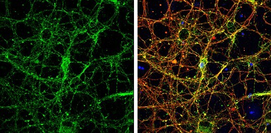
- Immunocytochemistry-Immunofluorescence analysis of HOMER1 was performed in DIV9 rat E18 primary cortical neurons fixed in 4% paraformaldehyde at RT for 15 min. Green: HOMER1 Polyclonal Antibody (Product # PA5-21487) diluted at 1:500. Red: beta Tubulin 3/ Tuj1, stained by beta Tubulin 3/ Tuj1 antibody. Blue: Fluoroshield with DAPI.
- Submitted by
- Invitrogen Antibodies (provider)
- Main image

- Experimental details
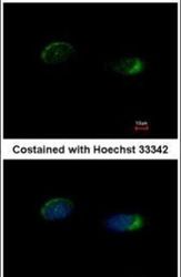
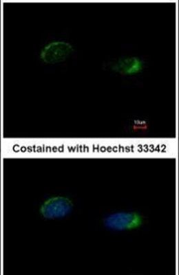
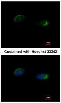
- Immunofluorescence analysis of methanol-fixed HeLa, using Homer antibody (Product # PA5-21487) at 1:500 dilution.
- Submitted by
- Invitrogen Antibodies (provider)
- Main image

- Experimental details
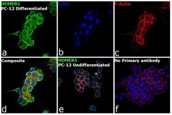
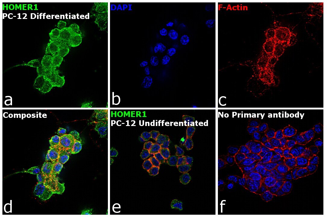
- Immunofluorescence analysis of HOMER1 was performed using 90% confluent log phase PC-12 cells differentiated to Neuronal cells. The cells were fixed with 4% paraformaldehyde for 10 minutes, permeabilized with 0.1% Triton™ X-100 for 15 minutes, and blocked with 1% BSA for 1 hour at room temperature. The cells were labeled with HOMER1 Rabbit Polyclonal Antibody (Product # PA5-21487) at 5 microgram/mL in 0.1% BSA, incubated at 4 degree Celsius overnight and then labeled with Goat anti-Rabbit IgG (H+L) Superclonal™ Secondary Antibody, Alexa Fluor® 488 conjugate (Product # A27034) at a dilution of 1:2000 for 45 minutes at room temperature (Panel a: green). Nuclei (Panel b: blue) were stained with ProLong™ Diamond Antifade Mountant with DAPI (Product # P36962). F-actin (Panel c: red) was stained with Rhodamine Phalloidin (Product # R415, 1:300). Panel d represents the merged image showing Cytoplasmic localization. Panel e shows undifferentiated cells with reduced signal. Panel f represents control cells with no primary antibody to assess background. The images were captured at 60X magnification.

- Submitted by
- Invitrogen Antibodies (provider)
- Main image

- Experimental details
- Immunofluorescence analysis of methanol-fixed HeLa, using Homer antibody (Product # PA5-21487) at 1:500 dilution.

- Submitted by
- Invitrogen Antibodies (provider)
- Main image
- Experimental details
- Immunofluorescence analysis of HOMER1 was performed using 90% confluent log phase PC-12 cells differentiated to Neuronal cells. The cells were fixed with 4% paraformaldehyde for 10 minutes, permeabilized with 0.1% Triton™ X-100 for 15 minutes, and blocked with 1% BSA for 1 hour at room temperature. The cells were labeled with HOMER1 Rabbit Polyclonal Antibody (Product # PA5-21487) at 5 microgram/mL in 0.1% BSA, incubated at 4 degree Celsius overnight and then labeled with Goat anti-Rabbit IgG (Heavy Chain) Superclonal™ Secondary Antibody, Alexa Fluor® 488 conjugate (Product # A27034) at a dilution of 1:2000 for 45 minutes at room temperature (Panel a: green). Nuclei (Panel b: blue) were stained with ProLong™ Diamond Antifade Mountant with DAPI (Product # P36962). F-actin (Panel c: red) was stained with Rhodamine Phalloidin (Product # R415, 1:300). Panel d represents the merged image showing Cytoplasmic localization. Panel e shows undifferentiated cells with reduced signal. Panel f represents control cells with no primary antibody to assess background. The images were captured at 60X magnification.
- Submitted by
- Invitrogen Antibodies (provider)
- Main image

- Experimental details
- Immunocytochemistry-Immunofluorescence analysis of HOMER1 was performed in DIV9 rat E18 primary cortical neurons fixed in 4% paraformaldehyde at RT for 15 min. Green: HOMER1 Polyclonal Antibody (Product # PA5-21487) diluted at 1:500. Red: beta Tubulin 3/ Tuj1, stained by beta Tubulin 3/ Tuj1 antibody. Blue: Fluoroshield with DAPI.

Supportive validation
- Submitted by
- Invitrogen Antibodies (provider)
- Main image
- Experimental details
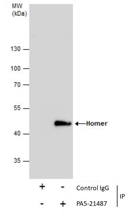
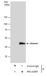
- Immunoprecipitation of Homer was performed in IMR32 whole cell extracts using 5 µg of HOMER1 Polyclonal Antibody (Product # PA5-21487). Samples were transferred to a membrane and probed with HOMER1 Polyclonal Antibody as a primary antibody and an HRP-conjugated anti-Rabbit IgG was used as a secondary antibody.

Supportive validation
- Submitted by
- Invitrogen Antibodies (provider)
- Main image
- Experimental details
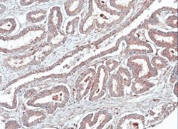
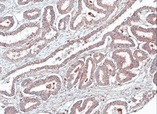
- Immunohistochemical analysis of paraffin-embedded human ovarian cancer, using HOMER1 (Product # PA5-21487) antibody at 1:100 dilution.
- Submitted by
- Invitrogen Antibodies (provider)
- Main image
- Experimental details
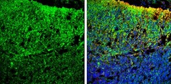
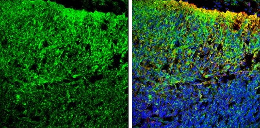
- Immunohistochemistry (Frozen) analysis of HOMER1 was performed in frozen sectioned E13.5 Rat brain tissue using HOMER1 Polyclonal Antibody (Product # PA5-21487) at a dilution of 1:250 (Green). Red: beta Tubulin 3/ TUJ1, a mature neuron marker, stained by beta Tubulin 3/ TUJ1 antibody diluted at 1:500. Blue: Fluoroshield with DAPI.
Supportive validation
- Submitted by
- Invitrogen Antibodies (provider)
- Main image

- Experimental details
- Immunoprecipitation of Homer was performed in IMR32 whole cell extracts using 5 µg of HOMER1 Polyclonal Antibody (Product # PA5-21487). Samples were transferred to a membrane and probed with HOMER1 Polyclonal Antibody as a primary antibody and an HRP-conjugated anti-Rabbit IgG was used as a secondary antibody.