Explore
 Validate
 Learn
LearnMAB91662-100
antibody from Novus Biologicals
Targeting: SERPINH1
CBP1, CBP2, colligen, HSP47, SERPINH2
 Western blot
Antibody data
- Antibody Data
- Antigen structure
- References [0]
- Comments [0]
- Validations
- Western blot [2]
- Immunohistochemistry [1]
Submit
Validation data
Reference
Comment
Report error
- Product number
- MAB91662-100 - Provider product page

- Provider
- Novus Biologicals
- Product name
- Mouse Monoclonal Hsp47 Antibody
- Antibody type
- Monoclonal
- Description
- Protein A or G purified from hybridoma culture supernatant. Detects human HSP47 in direct ELISAs and Western blots. In Western blots, no cross-reactivity with mouse HSP47 is observed.
- Reactivity
- Human
- Host
- Mouse
- Conjugate
- Unconjugated
- Isotype
- IgG
- Vial size
- 100 ug
- Storage
- Use a manual defrost freezer and avoid repeated freeze-thaw cycles. 12 months from date of receipt, -20 to -70 degreesC as supplied. 1 month, 2 to 8 degreesC under sterile conditions after reconstitution. 6 months, -20 to -70 degreesC under sterile conditions after reconstitution.
No comments: Submit comment
Supportive validation
- Submitted by
- Novus Biologicals (provider)
- Main image

- Experimental details
- Detection of Human HSP47 by Western Blot. Western blot shows lysates of HeLa human cervical epithelial carcinoma cell line, HepG2 human hepatocellular carcinoma cell line, A549 human lung carcinoma cell line, and JAR human choriocarcinoma cell line. PVDF membrane was probed with 0.1 µg/mL of Mouse Anti-Human HSP47 Monoclonal Antibody (Catalog # MAB91662) followed by HRP-conjugated Anti-Mouse IgG Secondary Antibody (Catalog # HAF018). A specific band was detected for HSP47 at approximately 47 kDa (as indicated). This experiment was conducted under reducing conditions and using Immunoblot Buffer Group 1.

- Submitted by
- Novus Biologicals (provider)
- Main image

- Experimental details
- Detection of Human HSP47 by Simple WesternTM. Simple Western lane view shows lysates of HeLa human cervical epithelial carcinoma cell line and HepG2 human hepatocellular carcinoma cell line, loaded at 0.2 mg/mL. A specific band was detected for HSP47 at approximately 57 kDa (as indicated) using 1 µg/mL of Mouse Anti-Human HSP47 Monoclonal Antibody (Catalog # MAB91662) . This experiment was conducted under reducing conditions and using the 12-230 kDa separation system.

Supportive validation
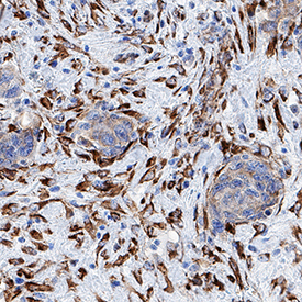
- Submitted by
- Novus Biologicals (provider)
- Main image
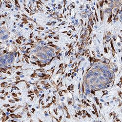
- Experimental details
- HSP47 in Human Breast Cancer Tissue. HSP47 was detected in immersion fixed paraffin-embedded sections of human breast cancer tissue using Mouse Anti-Human HSP47 Monoclonal Antibody (Catalog # MAB91662) at 25 µg/mL overnight at 4 °C. Tissue was stained using the Anti-Mouse HRP-DAB Cell & Tissue Staining Kit (brown; Catalog # CTS002) and counterstained with hematoxylin (blue). Specific staining was localized to cytoplasm. View our protocol for Chromogenic IHC Staining of Paraffin-embedded Tissue Sections.