Explore
 Validate
 Learn
LearnPA5-27832
antibody from Invitrogen Antibodies
Targeting: SERPINH1
CBP1, CBP2, colligen, HSP47, SERPINH2
 Western blot
 Immunocytochemistry
Antibody data
- Antibody Data
- Antigen structure
- References [4]
- Comments [0]
- Validations
- Immunocytochemistry [1]
- Immunohistochemistry [2]
- Other assay [1]
Submit
Validation data
Reference
Comment
Report error
- Product number
- PA5-27832 - Provider product page

- Provider
- Invitrogen Antibodies
- Product name
- SERPINH1 Polyclonal Antibody
- Antibody type
- Polyclonal
- Antigen
- Recombinant full-length protein
- Description
- Recommended positive controls: NIH-3T3, A431, HeLa, HepG2, U87-MG, SK-N-SH, IMR32, SK-N-AS. Predicted reactivity: Mouse (97%), Rat (97%), Dog (98%), Pig (96%), Chicken (88%), Sheep (96%), Rhesus Monkey (100%), Chimpanzee (100%), Bovine (96%). Store product as a concentrated solution. Centrifuge briefly prior to opening the vial.
- Reactivity
- Human, Mouse
- Host
- Rabbit
- Isotype
- IgG
- Vial size
- 100 μL
- Concentration
- 0.37 mg/mL
- Storage
- Store at 4°C short term. For long term storage, store at -20°C, avoiding freeze/thaw cycles.
Submitted references Small molecule inhibitor of HSP47 prevents pro-fibrotic mechanisms of fibroblasts in vitro.
Conditional deletion of Nedd4-2 in lung epithelial cells causes progressive pulmonary fibrosis in adult mice.
Detection of RAGE expression and its application to diabetic wound age estimation.
Activation of α7nAChR Promotes Diabetic Wound Healing by Suppressing AGE-Induced TNF-α Production.
Miyamura T, Sakamoto N, Kakugawa T, Taniguchi H, Akiyama Y, Okuno D, Moriyama S, Hara A, Kido T, Ishimoto H, Yamaguchi H, Miyazaki T, Obase Y, Ishimatsu Y, Tanaka Y, Mukae H
Biochemical and biophysical research communications 2020 Sep 24;530(3):561-565
Biochemical and biophysical research communications 2020 Sep 24;530(3):561-565
Conditional deletion of Nedd4-2 in lung epithelial cells causes progressive pulmonary fibrosis in adult mice.
Duerr J, Leitz DHW, Szczygiel M, Dvornikov D, Fraumann SG, Kreutz C, Zadora PK, Seyhan Agircan A, Konietzke P, Engelmann TA, Hegermann J, Mulugeta S, Kawabe H, Knudsen L, Ochs M, Rotin D, Muley T, Kreuter M, Herth FJF, Wielpütz MO, Beers MF, Klingmüller U, Mall MA
Nature communications 2020 Apr 24;11(1):2012
Nature communications 2020 Apr 24;11(1):2012
Detection of RAGE expression and its application to diabetic wound age estimation.
Ji XY, Chen Y, Ye GH, Dong MW, Lin KZ, Han JG, Feng XP, Li XB, Yu LS, Fan YY
International journal of legal medicine 2017 May;131(3):691-698
International journal of legal medicine 2017 May;131(3):691-698
Activation of α7nAChR Promotes Diabetic Wound Healing by Suppressing AGE-Induced TNF-α Production.
Dong MW, Li M, Chen J, Fu TT, Lin KZ, Ye GH, Han JG, Feng XP, Li XB, Yu LS, Fan YY
Inflammation 2016 Apr;39(2):687-99
Inflammation 2016 Apr;39(2):687-99
No comments: Submit comment
Supportive validation
- Submitted by
- Invitrogen Antibodies (provider)
- Main image
- Experimental details
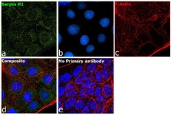
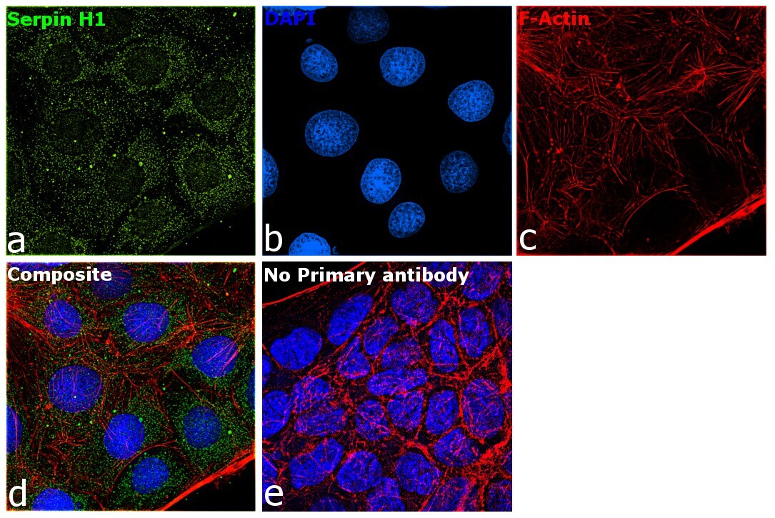
- Immunofluorescence analysis of Serpin H1 was performed using 70% confluent log phase Caco-2 cells. The cells were fixed with 4% paraformaldehyde for 5 minutes, permeabilized with 0.1% Triton™ X-100 for 10 minutes, and blocked with 2% BSA for 45 minutes at room temperature. The cells were labeled with SERPINH1 Polyclonal Antibody (Product # PA5-27832) at 1:100 dilution in 0.1% BSA, incubated at 4 degree celsius overnight and then labeled with Donkey anti-Rabbit IgG (H+L) Highly Cross-Adsorbed Secondary Antibody, Alexa Fluor Plus 488 (Product # A32790), (1:2000 dilution), for 45 minutes at room temperature (Panel a: Green). Nuclei (Panel b:Blue) were stained with ProLong™ Diamond Antifade Mountant with DAPI (Product # P36962). F-actin (Panel c: Red) was stained with Rhodamine Phalloidin (Product # R415, 1:300). Panel d represents the merged image showing cytoplasmic (endoplasmic reticulum) localization. Panel e represents control cells with no primary antibody to assess background. The images were captured at 60X magnification.
Supportive validation
- Submitted by
- Invitrogen Antibodies (provider)
- Main image
- Experimental details
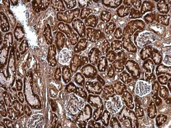
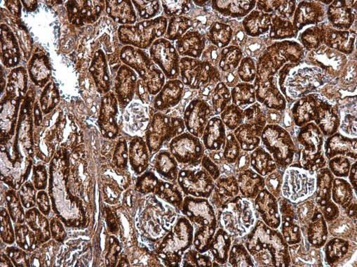
- SERPINH1 Polyclonal Antibody detects HSP47 protein at cytosol on mouse kidney by immunohistochemical analysis. Sample: Paraffin-embedded mouse kidney. SERPINH1 Polyclonal Antibody (Product # PA5-27832) dilution: 1:500. Antigen Retrieval: EDTA based buffer, pH 8.0, 15 min.
- Submitted by
- Invitrogen Antibodies (provider)
- Main image

- Experimental details
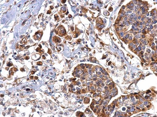
- SERPINH1 Polyclonal Antibody detects HSP47 protein at cytosol on human breast carcinoma by immunohistochemical analysis. Sample: Paraffin-embedded human breast carcinoma. SERPINH1 Polyclonal Antibody (Product # PA5-27832) dilution: 1:500. Antigen Retrieval: EDTA based buffer, pH 8.0, 15 min.
Supportive validation
- Submitted by
- Invitrogen Antibodies (provider)
- Main image
- Experimental details
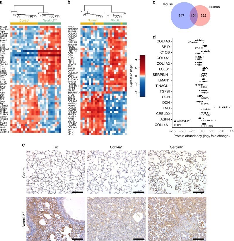
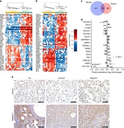
- Fig. 7 Comparison of proteomic signatures of pulmonary fibrosis in conditional Nedd4-2 -/- mice and patients with IPF. a , b Heatmap of differentially regulated matrisome annotated proteins in lungs from conditional Nedd4-2 -/- mice ( n = 11) and control mice ( n = 13) induced with doxycycline for 3 months ( a ), and in lung tissues from IPF patients and controls ( n = 11/group) ( b ). c Venn-diagram showing proportion of unique and common differentially regulated proteins in lungs of conditional Nedd4-2 - /- mice and IPF patients. d Summary of differentially regulated matrisome proteins found in conditional Nedd4-2 - /- mice and human IPF samples. e Micrographs of representative lung sections from conditional Nedd4-2 - /- mice and littermate controls stained with anti-Tnc, anti-Col14a1, and anti-Serpinh1 antibodies ( n = 6/group). Scale bars, 100 um.