 Explore
 Validate
 Learn
Learn Western blot
 Immunohistochemistry
Antibody data
- Antibody Data
- Antigen structure
- References [2]
- Comments [0]
- Validations
- Immunohistochemistry [4]
- Other assay [3]
Submit
Validation data
Reference
Comment
Report error
- Product number
- PA5-79199 - Provider product page

- Provider
- Invitrogen Antibodies
- Product name
- HuD Polyclonal Antibody
- Antibody type
- Polyclonal
- Antigen
- Synthetic peptide
- Description
- Reconstitute with 0.2 mL of distilled water to yield a concentration of 500 µg/mL. Positive Control - WB: rat brain tissue, mouse brain tissue, U87-MG whole cell. IHC: mouse brain tissue, rat brain tissue, human meningeoma tissue.
- Reactivity
- Human, Mouse, Rat
- Host
- Rabbit
- Isotype
- IgG
- Vial size
- 100 μg
- Concentration
- 500 μg/mL
- Storage
- -20°C
Submitted references Myenteric Neurons Do Not Replicate in Small Intestine Under Normal Physiological Conditions in Adult Mouse.
The ephrin receptor EphB2 regulates the connectivity and activity of enteric neurons.
Virtanen H, Garton DR, Andressoo JO
Cellular and molecular gastroenterology and hepatology 2022;14(1):27-34
Cellular and molecular gastroenterology and hepatology 2022;14(1):27-34
The ephrin receptor EphB2 regulates the connectivity and activity of enteric neurons.
Bodin R, Paillé V, Oullier T, Durand T, Aubert P, Le Berre-Scoul C, Hulin P, Neunlist M, Cissé M
The Journal of biological chemistry 2021 Nov;297(5):101300
The Journal of biological chemistry 2021 Nov;297(5):101300
No comments: Submit comment
Supportive validation
- Submitted by
- Invitrogen Antibodies (provider)
- Main image
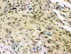
- Experimental details
- Immunohistochemistry analysis of HuD on paraffin-embedded human meningioma tissue. Sample was incubated with HuD polyclonal antibody (Product# PA5-79199).
- Submitted by
- Invitrogen Antibodies (provider)
- Main image
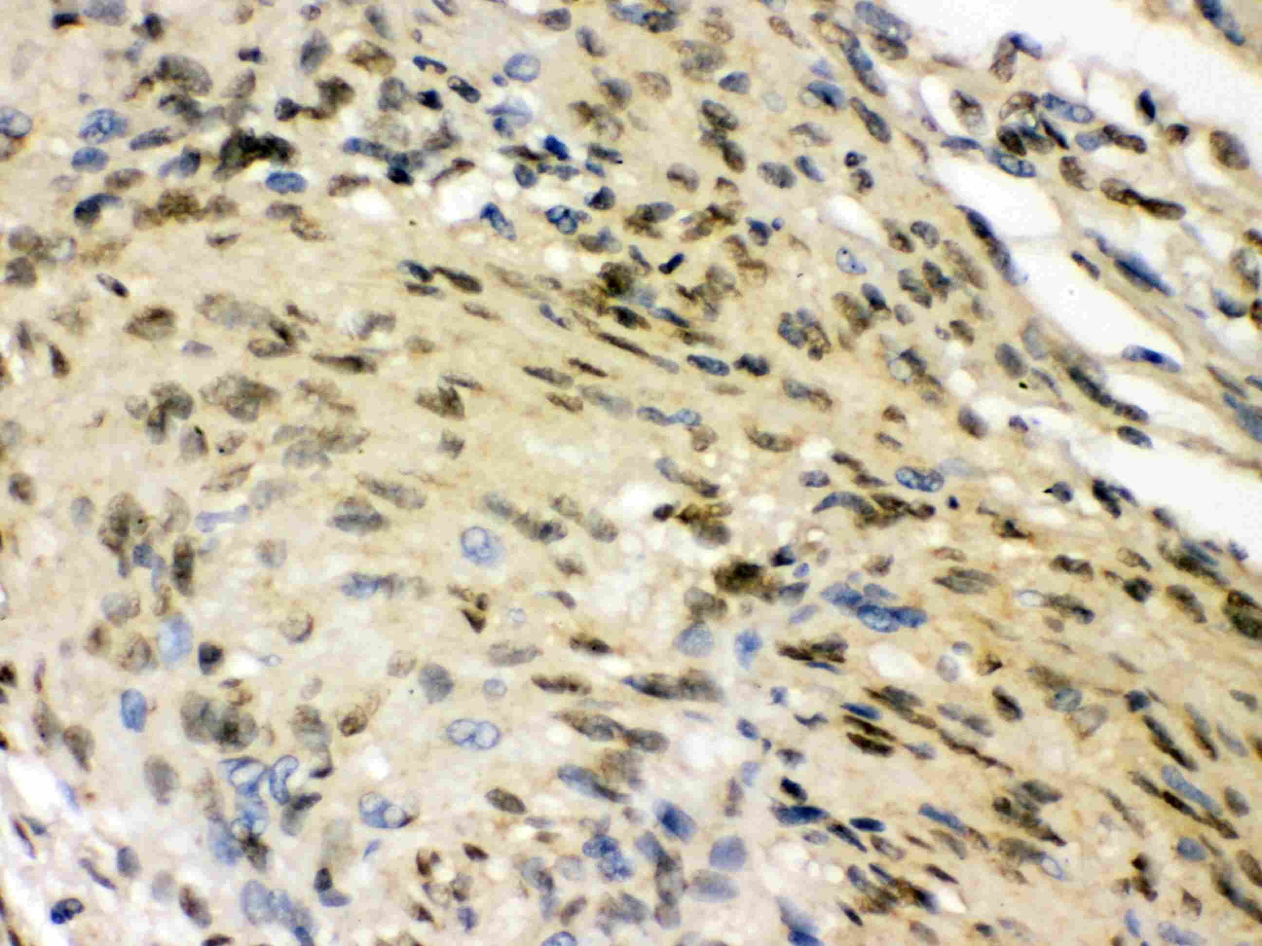
- Experimental details
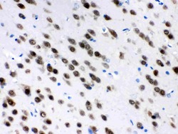
- Immunohistochemistry analysis of HuD on paraffin-embedded mouse brain tissue. Sample was incubated with HuD polyclonal antibody (Product# PA5-79199).
- Submitted by
- Invitrogen Antibodies (provider)
- Main image
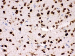
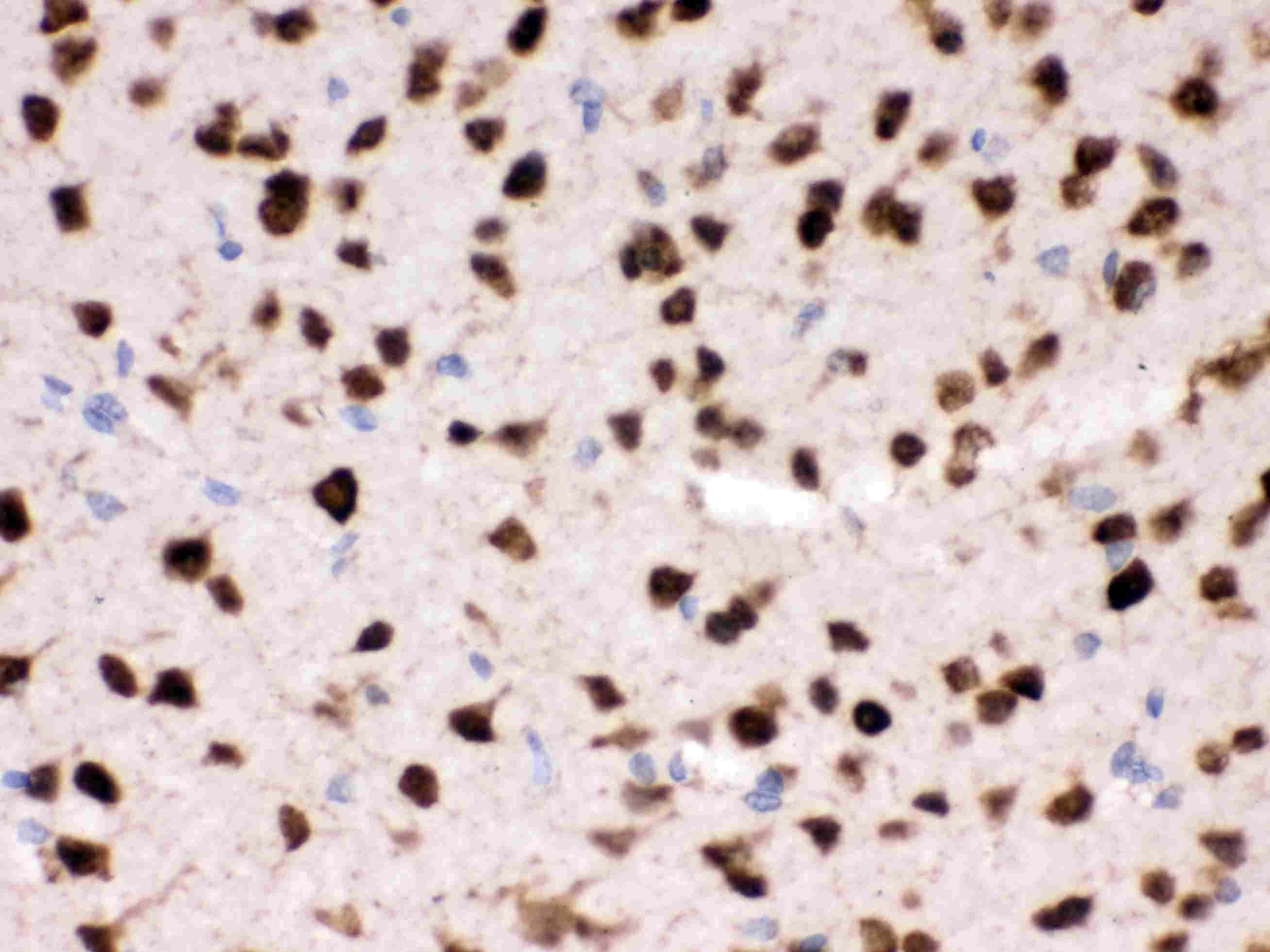
- Experimental details
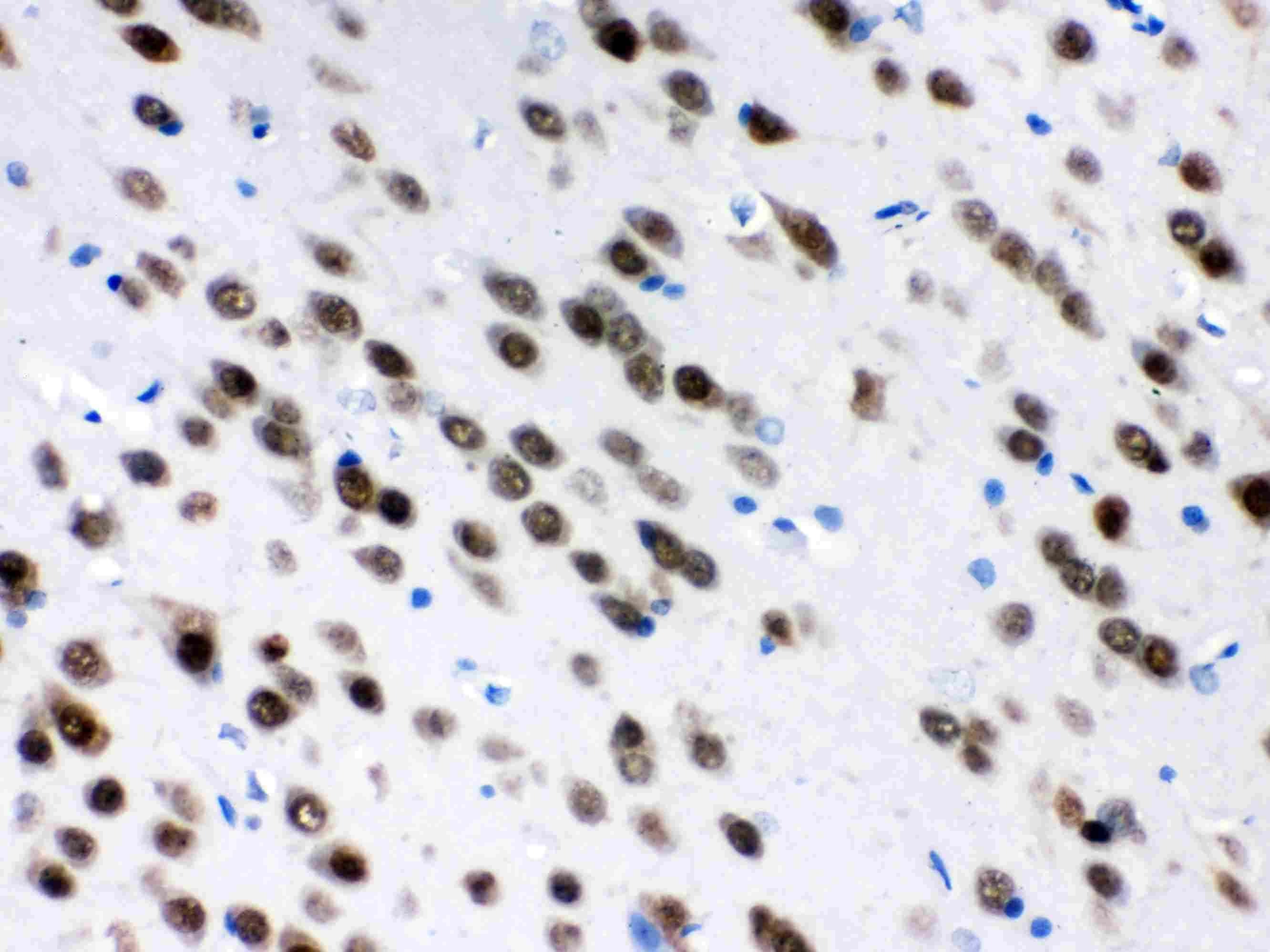
- Immunohistochemistry analysis of HuD on paraffin-embedded rat brain tissue. Sample was incubated with HuD polyclonal antibody (Product# PA5-79199).
- Submitted by
- Invitrogen Antibodies (provider)
- Main image

- Experimental details
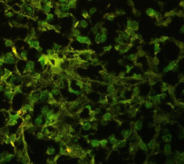
- Immunohistochemical analysis of ELAVL4 in paraffin-embedded section of human glioma tissues. Heat mediated antigen retrieval was performed in citrate buffer (pH6, epitope retrieval solution ) for 20 mins. The tissue section was blocked with 10% goat serum. The tissue section was then incubated with 1μg/mL rabbit anti-ELAVL4 antibody (Product # PA5-79199) overnight at 4°C. DyLight®488 Conjugated Goat Anti-Rabbit IgG was used as secondary antibody at 1:100 dilution and incubated for 30 minutes at 37°C. Visualize using a fluorescence microscope and filter sets appropriate for the label used.
Supportive validation
- Submitted by
- Invitrogen Antibodies (provider)
- Main image
- Experimental details
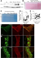
- Figure 1 EphB2 expression in different segments of the rat GI tract. A - C , EphB2 expression was examined by western blot analysis of full-thickness tissue sections from indicated regions of the rat GI tract. Pgp9.5 was used as a loading control ( A ). Note that pgp9.5 is expressed at higher levels in the distal colon. Furthermore, we observed the apparition of putative EphB2-derived bands in the colon that might be degradation products. Ponceau S ( B ) and Coomassie ( C ) stainings are provided as addition to the traditional loading control. Although equal amount of proteins was loaded for analysis, those stainings show the variability in protein migration profil and relative protein abundance between intestinal segments. Each data point represents the relative adjusted volume of protein band intensity measured by using a rectangle tool of constant volume on each lane of the WB membrane. Data are represented as relative values of protein density in arbitrary units. D , densitometric quantitation of western blot signals revealed that EphB2 is present in all segments of the gut (n = 12 sections per segment from 12 rats; jej., jejunum; prox. colon, proximal colon; dist. colon, distal colon; n = 12 sections per segment from 12 animals, F (3, 44) = 18.91, p = 0.00000005, ANOVA; Tukey's post-hoc test; ** p < 0.001, *** p < 0.0001). E , quantitative RT-PCR analysis of EphB2 mRNA levels in full-thickness tissue sections in the GI tract of adult rats. Note that there is no strict pr

- Submitted by
- Invitrogen Antibodies (provider)
- Main image
- Experimental details
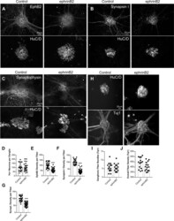
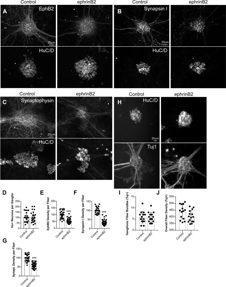
- Figure 5 Chronic activation with ephrinB2 reduces levels of EphB2 and synaptic-associated proteins in ENS cultures. A - C , primary cultures of E15 rat intestines were grown on glass coverslips and fully matured into an enteric-like neuronal network with ganglia interconnected with neural fibers at 12 DIV. Cultures were then treated with control-Fc (Control) or ephrinB2-Fc ligand (ephrinB2) for 5 consecutive days. Cultures were fixed and double-immunostained for EphB2 ( A ), synapsin I ( B ), synaptophysin ( C ) and pan-neuronal marker HuC/D ( A - C ). Images represent ganglia depicting neuronal cell bodies and processes. D - F , quantitations of images represented in ( A - C ) for HuC/D-positive neurons ( D ), and neuronal fiber density of EphB2 ( E ), synapsin I ( F ) or synaptophysin ( G ). Note that chronic exposure of neurons to ephrinB2 decreased EphB2 expression within neuronal processes without affecting overall neuronal density. Synapsin I and synaptophysin were reduced within neuronal processes. H , images represent ganglia depicting neuronal cell bodies (HuC/D staining) and neuronal processes (Tuj-1 staining). I and J , quantitations of images represented in ( H ) for the number of neuronal processes/ganglionic fiber bundles ( I ) and overall fiber density ( J ). (n = 10-15 ganglia per condition from at least three independent cultures, unpaired t test). *** p
- Submitted by
- Invitrogen Antibodies (provider)
- Main image
- Experimental details
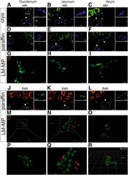
- Analysis of ENS proliferation in the small intestine. ( A-C ) Longitudinal cryosections and ( D-F ) paraffin sections of the small intestine were immunostained for IdU ( green ) and for HuD ( blue ) to reveal enteric neurons that had undergone replication. Cells in the epithelium show signal for IdU as expected. ( A-F ) Myenteric ganglia are indicated with white arrowheads and submucosal ganglia with yellow arrowheads . No double-positive HuD+/IdU+ enteric neurons were detected (n = 300 ganglia analyzed in n = 3 animals, age = 24 weeks). Dashed rectangle indicates magnified area next to the panel. Scale bar, 20 mum, 10 mum on insets. ( G-I ) Analysis of LM-MPs from IdU-labelled animals revealed no double-positive HuD+/IdU+ enteric neurons (n = 3790 neurons analyzed in n = 3 animals, age = 21 weeks). Scale bar, 20 mum. ( J-L ) Analysis of longitudinal paraffin sections from the small intestine of EdU-labelled animals, HuD ( green ) and EdU ( red ). Cells in the epithelium show signal for EdU as expected. No double-positive HuD+/EdU+ enteric neurons were detected (n = 451 ganglia counted in n = 3 animals, age = 17 weeks). Myenteric ganglia are indicated with white arrowheads and submucosal ganglia with yellow arrowheads . Dashed rectangle indicates magnified area next to the panel. Scale bar, 20 mum, 10 mum on insets. ( M-O ) Analysis of LM-MPs from EdU-labelled animals (n = 1474 neurons analyzed in n = 4 animals, age = 8-10 weeks) revealed no double-positive HuD+/EdU+ enteric
