Explore
 Validate
 Learn
Learn Western blot
 Immunocytochemistry
Antibody data
- Antibody Data
- Antigen structure
- References [6]
- Comments [0]
- Validations
- Immunocytochemistry [1]
- Flow cytometry [2]
Submit
Validation data
Reference
Comment
Report error
- Product number
- 14-1249-82 - Provider product page

- Provider
- Invitrogen Antibodies
- Product name
- CD124 Monoclonal Antibody (X2/45-12), eBioscience™
- Antibody type
- Monoclonal
- Antigen
- Other
- Description
- Description: The monoclonal antibody X2/45-12 reacts with human CD124, which is also known as the IL-4R alpha chain. CD124 can associate with the common gamma chain to form the type I IL-4R or with IL-13Ralpha1 to form the type II IL-4R. While both receptor types can bind IL-4, only the type II receptor can associate with IL-13. The type I IL-4 receptor is found on T, B, NK, mast cells, basophils, and macrophages. The type II IL-4 receptor is found on many non-hematopoietic cells and macrophages. CD124 contains an ITIM that associates with and activates STAT6. IL-4 is responsible for the generation of high-affinity IgE antibodies and driving the polarization of naive Th0 cells into Th2 cells. In mucosal barriers, the cytokine is involved in the recruitment of innate and adaptive immune effector cells and mucous overproduction. Finally, IL-4R also exists as a soluble form that serves primarily as a carrier protein for IL-4 that increases its half-life. The X2/45 antibody has been reported to have antagonist activity. Applications Reported: This X2/45-12 antibody has been reported for use in flow cytometric analysis, and western blotting. (Please use Functional Grade purified X2/45-12, Product # 16-1249-82, in functional assays). Applications Tested: This X2/45-12 antibody has been tested by flow cytometric analysis of normal human peripheral blood cells. This can be used at less than or equal to 1 µg per test. A test is defined as the amount (µg) of antibody that will stain a cell sample in a final volume of 100 µL. Cell number should be determined empirically but can range from 10^5 to 10^8 cells/test. It is recommended that the antibody be carefully titrated for optimal performance in the assay of interest. Purity: Greater than 90%, as determined by SDS-PAGE. Aggregation: Less than 10%, as determined by HPLC. Filtration: 0.2 µm post-manufacturing filtered.
- Reactivity
- Human
- Host
- Mouse
- Isotype
- IgG
- Antibody clone number
- X2/45-12
- Vial size
- 100 μg
- Concentration
- 0.5 mg/mL
- Storage
- 4°C
Submitted references Interleukin-4- and interleukin-13-mediated alternatively activated macrophages: roles in homeostasis and disease.
Differences in expression, affinity, and function of soluble (s)IL-4Ralpha and sIL-13Ralpha2 suggest opposite effects on allergic responses.
Tissue-specific function of lymph node fibroblastic reticulum cells.
IL-4 receptor alpha is an important modulator of IL-4 and IL-13 receptor binding: implications for the development of therapeutic targets.
Interleukin-13 overexpression by tax transactivation: a potential autocrine stimulus in human T-cell leukemia virus-infected lymphocytes.
Design of human interleukin-4 antagonists inhibiting interleukin-4-dependent and interleukin-13-dependent responses in T-cells and B-cells with high efficiency.
Van Dyken SJ, Locksley RM
Annual review of immunology 2013;31:317-43
Annual review of immunology 2013;31:317-43
Differences in expression, affinity, and function of soluble (s)IL-4Ralpha and sIL-13Ralpha2 suggest opposite effects on allergic responses.
Khodoun M, Lewis CC, Yang JQ, Orekov T, Potter C, Wynn T, Mentink-Kane M, Hershey GK, Wills-Karp M, Finkelman FD
Journal of immunology (Baltimore, Md. : 1950) 2007 Nov 15;179(10):6429-38
Journal of immunology (Baltimore, Md. : 1950) 2007 Nov 15;179(10):6429-38
Tissue-specific function of lymph node fibroblastic reticulum cells.
Vega F, Coombes KR, Thomazy VA, Patel K, Lang W, Jones D
Pathobiology : journal of immunopathology, molecular and cellular biology 2006;73(2):71-81
Pathobiology : journal of immunopathology, molecular and cellular biology 2006;73(2):71-81
IL-4 receptor alpha is an important modulator of IL-4 and IL-13 receptor binding: implications for the development of therapeutic targets.
Andrews AL, Holloway JW, Holgate ST, Davies DE
Journal of immunology (Baltimore, Md. : 1950) 2006 Jun 15;176(12):7456-61
Journal of immunology (Baltimore, Md. : 1950) 2006 Jun 15;176(12):7456-61
Interleukin-13 overexpression by tax transactivation: a potential autocrine stimulus in human T-cell leukemia virus-infected lymphocytes.
Wäldele K, Schneider G, Ruckes T, Grassmann R
Journal of virology 2004 Jun;78(12):6081-90
Journal of virology 2004 Jun;78(12):6081-90
Design of human interleukin-4 antagonists inhibiting interleukin-4-dependent and interleukin-13-dependent responses in T-cells and B-cells with high efficiency.
Tony HP, Shen BJ, Reusch P, Sebald W
European journal of biochemistry 1994 Oct 15;225(2):659-65
European journal of biochemistry 1994 Oct 15;225(2):659-65
No comments: Submit comment
Supportive validation
- Submitted by
- Invitrogen Antibodies (provider)
- Main image
- Experimental details
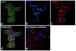
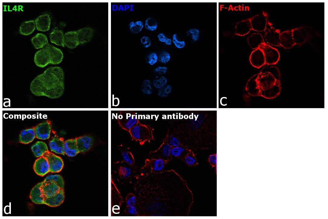
- Immunofluorescence analysis of Interleukin-4 receptor subunit alpha was performed using 70% confluent log phase PANC-1 cells. The cells were fixed with 4% paraformaldehyde for 10 minutes, permeabilized with 0.1% Triton™ X-100 for 15 minutes, and blocked with 2% BSA for 45 minutes at room temperature. The cells were labeled with CD124 Monoclonal Antibody (X2/45-12), eBioscience™ (Product # 14-1249-82) at 1:100 dilution in 0.1% BSA, incubated at 4 degrees celsius overnight, and then labeled with Donkey anti-Mouse IgG (H+L) Highly Cross-Adsorbed Secondary Antibody, Alexa Fluor Plus 488 (Product # A32766), (1:2000 dilution), for 45 minutes at room temperature (Panel a: Green). Nuclei (Panel b: Blue) were stained with ProLong™ Diamond Antifade Mountant with DAPI (Product # P36962). F-actin (Panel c: Red) was stained with Rhodamine Phalloidin (Product # R415, 1:300 dilution). Panel d represents the merged image showing membranous localization. Panel e represents control cells with no primary antibody to assess the background. The images were captured at 60X magnification.
Supportive validation
- Submitted by
- Invitrogen Antibodies (provider)
- Main image
- Experimental details
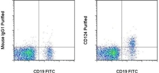
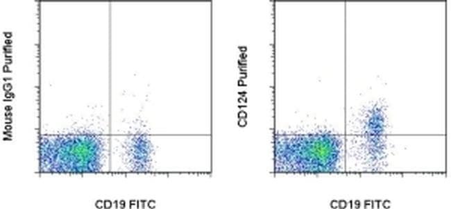
- Staining of normal human peripheral blood cells with Anti-Human CD19 FITC (Product # 11-0199-42) and 0.5 µg of Mouse IgG1 K Isotype Control Purified (Product # 14-4714-82) (left) or 0.5 µg of Anti-Human CD124 Purified (right) followed by Anti-Mouse IgG1 Biotin (Product # 13-4015-82) and Streptavidin APC (Product # 17-4314-82). Cells in the lymphocyte gate were used for analysis.
- Submitted by
- Invitrogen Antibodies (provider)
- Main image

- Experimental details
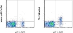
- Staining of normal human peripheral blood cells with Anti-Human CD19 FITC (Product # 11-0199-42) and 0.5 µg of Mouse IgG1 K Isotype Control Purified (Product # 14-4714-82) (left) or 0.5 µg of Anti-Human CD124 Purified (right) followed by Anti-Mouse IgG1 Biotin (Product # 13-4015-82) and Streptavidin APC (Product # 17-4314-82). Cells in the lymphocyte gate were used for analysis.