Explore
 Validate
 Learn
Learn Western blot
 Immunoprecipitation
Antibody data
- Antibody Data
- Antigen structure
- References [2]
- Comments [0]
- Validations
- Western blot [1]
- Immunocytochemistry [1]
- Other assay [3]
Submit
Validation data
Reference
Comment
Report error
- Product number
- PA5-19855 - Provider product page

- Provider
- Invitrogen Antibodies
- Product name
- IRAK1 Polyclonal Antibody
- Antibody type
- Polyclonal
- Antigen
- Synthetic peptide
- Description
- A suggested positive control is Hela cell lysate. PA5-19855 can be used with blocking peptide PEP-0001.
- Reactivity
- Human, Mouse, Rat
- Host
- Rabbit
- Isotype
- IgG
- Vial size
- 100 μg
- Concentration
- 1 mg/mL
- Storage
- Maintain refrigerated at 2-8°C for up to 3 months. For long term storage store at -20°C
Submitted references Human urine-derived stem cells protect against renal ischemia/reperfusion injury in a rat model via exosomal miR-146a-5p which targets IRAK1.
Estradiol/GPER affects the integrity of mammary duct-like structures in vitro.
Li X, Liao J, Su X, Li W, Bi Z, Wang J, Su Q, Huang H, Wei Y, Gao Y, Li J, Liu L, Wang C
Theranostics 2020;10(21):9561-9578
Theranostics 2020;10(21):9561-9578
Estradiol/GPER affects the integrity of mammary duct-like structures in vitro.
Deng Y, Miki Y, Nakanishi A
Scientific reports 2020 Jan 28;10(1):1386
Scientific reports 2020 Jan 28;10(1):1386
No comments: Submit comment
Supportive validation
- Submitted by
- Invitrogen Antibodies (provider)
- Main image
- Experimental details
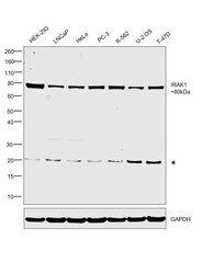
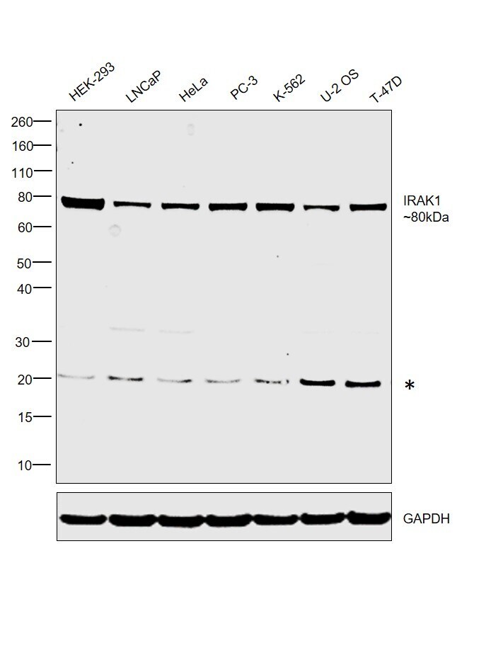
- Western blot was performed using Anti-IRAK1 Polyclonal Antibody (Product # PA5-19855) and an 80 kDa band corresponding to IRAK1 was observed across cell lines tested. Whole cell extracts (30 µg lysate) of HEK-293 (Lane 1), LNCaP (Lane 2), HeLa (Lane 3), PC-3 (Lane 4), K-562 (Lane 5), U-2 OS (Lane 6) and T-47D (Lane 7) were electrophoresed using Novex® NuPAGE® 4-12 % Bis-Tris gel (Product # NP0322BOX). Resolved proteins were then transferred onto a nitrocellulose membrane (Product # IB23001) by iBlot® 2 Dry Blotting System (Product # IB21001). The blot was probed with the primary antibody (1 µg/mL) and detected by chemiluminescence with Goat anti-Rabbit IgG (Heavy Chain), Superclonal™ Recombinant Secondary Antibody, HRP (Product # A27036, 1:4,000 dilution) using the iBright FL 1000 (Product # A32752). Chemiluminescent detection was performed using Novex® ECL Chemiluminescent Substrate Reagent Kit (Product # WP20005). An uncharacterized band (*) was observed at ~20 kDa.
Supportive validation
- Submitted by
- Invitrogen Antibodies (provider)
- Main image
- Experimental details
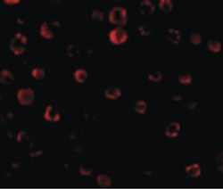
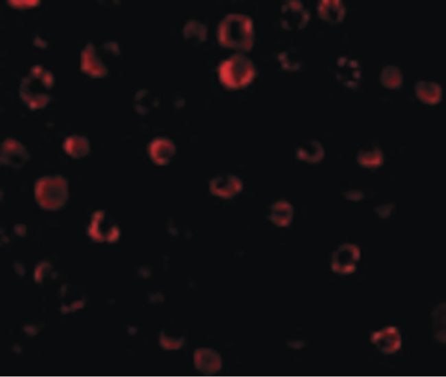
- Immunofluorescent analysis of HeLa cells using a IRAK polyclonal antibody (Product # PA5-19855) at a 20 µg/mL dilution.
Supportive validation
- Submitted by
- Invitrogen Antibodies (provider)
- Main image
- Experimental details
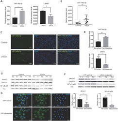
- Figure 6 USCs or USC-Exo upregulate miR-146a-5p expression, which targets the IRAK1 and NF-kappaB signaling in vivo and in vitro . Rat IRI was induced before treatment with or without USCs (annotated as USCs and control, respectively). ( A ) qRT-PCR analysis of the relative expression levels of miR-146a-5p ( P =0.0004) and IRAK1 ( P =0.0227). ( B ) qRT-PCR analysis of the relative expression levels of miR-146a-5p in rat serum exosomes on day 3 (n=10 in each group, P =0.0012). ( C ) FISH images of miR-146a-5p expression in kidney sections (scale bars = 100 um). ( D ) Western blot analysis of the protein levels of IRAK1 and nuclear NF-kappaB p65. n=5 in each group. GAPDH and H3 were used as loading controls, respectively. H/R injury was induced in HK2 cells in the absence or presence of USC-Exo (annotated as HK2 medium and HK2 medium+exosomes, respectively). ( E ) qRT-PCR analysis of the relative expression levels of miR-146a-5p ( P =0.0078) and IRAK1 ( P =0.0358). ( F ) Western blot assay of the protein levels of IRAK1 and nuclear NF-kappaB p65. n=3 in each group. GAPDH and H3 were used as loading controls, respectively. ( G ) Immunofluorescence analysis showed that NF-kappaB p65 in HK2 cells was transferred from the cytoplasm to the nucleus after H/R treatment, and this nuclear translocation of NF-kappaB p65 could be inhibited by USC-Exo treatment (scale bars = 50 um). Each experiment was repeated three times. Data represent the mean +- SEM. * P

- Submitted by
- Invitrogen Antibodies (provider)
- Main image

- Experimental details
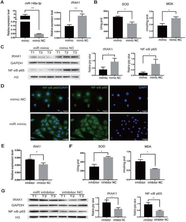
- Figure 7 miR-146a-5p mimic reduces oxidative stress and inhibits IRAK1 and NF-kappaB signaling in H/R-induced injury of HK2 cells. HK2 cells were transfected with miR-146a-5p mimic or its inhibitor before the H/R process. ( A ) qRT-PCR analysis of the relative expression levels of miR-146a-5p and IRAK1 in the mimic and mimic negative control (mimic NC) groups ( P
- Submitted by
- Invitrogen Antibodies (provider)
- Main image
- Experimental details
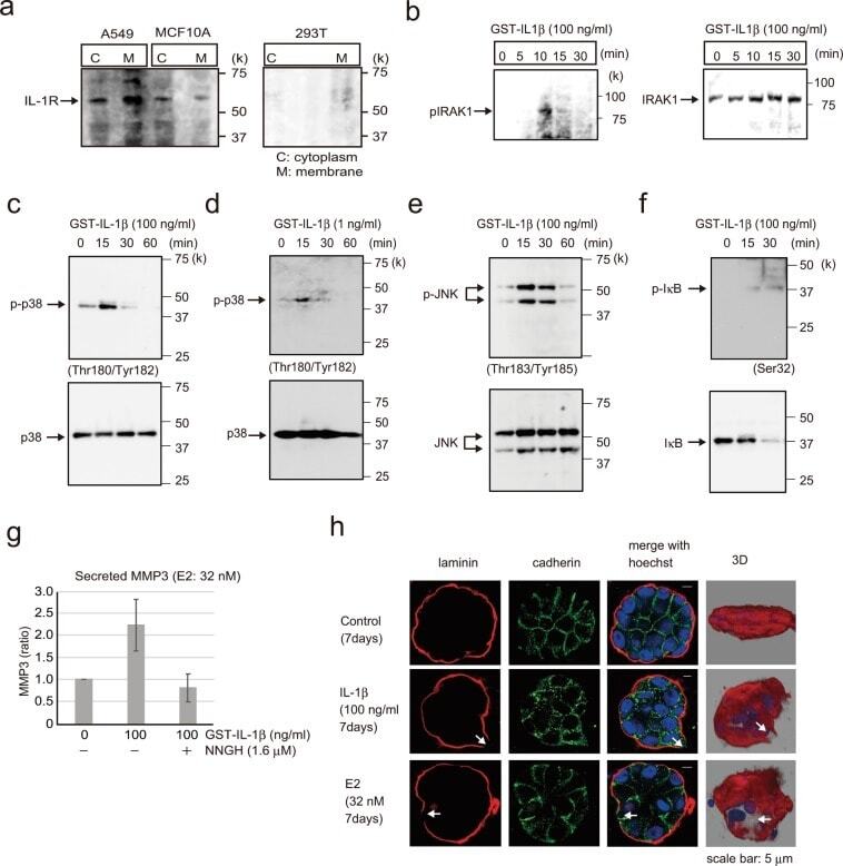
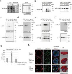
- Figure 5 Analysis of E2 signal transduction. ( a ) Western blotting of A549, MCF-10A, and 293T cell lysates showing IL-1R expression; cytoplasmic and membrane components of the cell lysates were separated. A549 cells were included as a positive control for IL-1R1 expression, and 293T cells were used as a negative control. ( b ) Western blotting of MCF-10A cells showing phospho-IRAK1 (left) and IRAK1 (right) following treatment with 100 ng/ml GST-IL-1beta for 0-30 min. ( c ) Western blotting of MCF-10A cells probed to examine phospho-p38(Thr180/Tyr182) and p38 after adding 100 ng/ml GST-IL-1beta for 0-60 min. ( d ) Western blotting of MCF-10A cells probed to examine phospho-p38 (Thr180/Tyr182) and p38 after adding 1 ng/ml GST-IL-1beta for 0-60 min. ( e ) Western blotting of MCF-10A cells showing phospho-JNK(Thr183/Tyr185) and JNK following treatment with 100 ng/ml GST-IL-1beta for 0-60 min. ( f ) Western blotting of MCF-10A cells showing phospho-IkB (Ser32) and IkB following treatment with 100 ng/ml GST-IL-1beta for 0-30 min. ( g ) MMP-3 activity assay of MCF-10A cells following treatment with 100 ng/ml GST-IL-1beta with or without 1.6 muM NNGH, which was used as an MMP-3 inhibitor. Four independent experiments were performed. Bars represent +/-SD. ( h ) Representative confocal images of MCF-10A cells in a 3D culture treated with 100 ng/ml GST-IL-1beta (second row) or E2 (32 nM, third row) or control (first row) for 7 days to examine the basement membrane via immunofluorescenc