 Explore
 Validate
 Learn
Learn Western blot
Antibody data
- Antibody Data
- Antigen structure
- References [11]
- Comments [0]
- Validations
- Western blot [2]
- Immunohistochemistry [2]
Submit
Validation data
Reference
Comment
Report error
- Product number
- AF1205 - Provider product page

- Provider
- Novus Biologicals
- Product name
- Rabbit Polyclonal JNK1/2/3 Antibody
- Antibody type
- Polyclonal
- Description
- Antigen and protein A Affinity-purified. Detects human, mouse and rat p46 and p54 JNK when dually phosphorylated at sites homologous to T183/Y185 of JNK1 and JNK2, and T221/Y223 of JNK3 in Western blots.
- Reactivity
- Human, Mouse, Rat
- Host
- Rabbit
- Conjugate
- Unconjugated
- Isotype
- IgG
- Vial size
- 50 ug
- Concentration
- LYOPH
- Storage
- Use a manual defrost freezer and avoid repeated freeze-thaw cycles. 12 months from date of receipt, -20 to -70 degreesC as supplied. 1 month, 2 to 8 degreesC under sterile conditions after reconstitution. 6 months, -20 to -70 degreesC under sterile conditions after reconstitution.
Submitted references Dysregulation of the splicing machinery is directly associated to aggressiveness of prostate cancer.
Effects of interleukin-17A in nucleus pulposus cells and its small-molecule inhibitors for intervertebral disc disease.
Mechanistic clues to the protective effect of chrysin against doxorubicin-induced cardiomyopathy: Plausible roles of p53, MAPK and AKT pathways.
WWOX sensitises ovarian cancer cells to paclitaxel via modulation of the ER stress response.
White-to-brown metabolic conversion of human adipocytes by JAK inhibition.
High glucose alters retinal astrocytes phenotype through increased production of inflammatory cytokines and oxidative stress.
A p38MAPK/MK2 signaling pathway leading to redox stress, cell death and ischemia/reperfusion injury.
Protective effect of the poly(ADP-ribose) polymerase inhibitor PJ34 on mitochondrial depolarization-mediated cell death in hepatocellular carcinoma cells involves attenuation of c-Jun N-terminal kinase-2 and protein kinase B/Akt activation.
ERK5 protein promotes, whereas MEK1 protein differentially regulates, the Toll-like receptor 2 protein-dependent activation of human endothelial cells and monocytes.
Oxidized {alpha}1-antitrypsin stimulates the release of monocyte chemotactic protein-1 from lung epithelial cells: potential role in emphysema.
Common dysregulation of Wnt/Frizzled receptor elements in human hepatocellular carcinoma.
Jiménez-Vacas JM, Herrero-Aguayo V, Montero-Hidalgo AJ, Gómez-Gómez E, Fuentes-Fayos AC, León-González AJ, Sáez-Martínez P, Alors-Pérez E, Pedraza-Arévalo S, González-Serrano T, Reyes O, Martínez-López A, Sánchez-Sánchez R, Ventura S, Yubero-Serrano EM, Requena-Tapia MJ, Castaño JP, Gahete MD, Luque RM
EBioMedicine 2020 Jan;51:102547
EBioMedicine 2020 Jan;51:102547
Effects of interleukin-17A in nucleus pulposus cells and its small-molecule inhibitors for intervertebral disc disease.
Suyama K, Sakai D, Hirayama N, Nakamura Y, Matsushita E, Terayama H, Qu N, Tanaka O, Sakabe K, Watanabe M
Journal of cellular and molecular medicine 2018 Nov;22(11):5539-5551
Journal of cellular and molecular medicine 2018 Nov;22(11):5539-5551
Mechanistic clues to the protective effect of chrysin against doxorubicin-induced cardiomyopathy: Plausible roles of p53, MAPK and AKT pathways.
Mantawy EM, Esmat A, El-Bakly WM, Salah ElDin RA, El-Demerdash E
Scientific reports 2017 Jul 6;7(1):4795
Scientific reports 2017 Jul 6;7(1):4795
WWOX sensitises ovarian cancer cells to paclitaxel via modulation of the ER stress response.
Janczar S, Nautiyal J, Xiao Y, Curry E, Sun M, Zanini E, Paige AJ, Gabra H
Cell death & disease 2017 Jul 27;8(7):e2955
Cell death & disease 2017 Jul 27;8(7):e2955
White-to-brown metabolic conversion of human adipocytes by JAK inhibition.
Moisan A, Lee YK, Zhang JD, Hudak CS, Meyer CA, Prummer M, Zoffmann S, Truong HH, Ebeling M, Kiialainen A, Gérard R, Xia F, Schinzel RT, Amrein KE, Cowan CA
Nature cell biology 2015 Jan;17(1):57-67
Nature cell biology 2015 Jan;17(1):57-67
High glucose alters retinal astrocytes phenotype through increased production of inflammatory cytokines and oxidative stress.
Shin ES, Huang Q, Gurel Z, Sorenson CM, Sheibani N
PloS one 2014;9(7):e103148
PloS one 2014;9(7):e103148
A p38MAPK/MK2 signaling pathway leading to redox stress, cell death and ischemia/reperfusion injury.
Ashraf MI, Ebner M, Wallner C, Haller M, Khalid S, Schwelberger H, Koziel K, Enthammer M, Hermann M, Sickinger S, Soleiman A, Steger C, Vallant S, Sucher R, Brandacher G, Santer P, Dragun D, Troppmair J
Cell communication and signaling : CCS 2014 Jan 14;12:6
Cell communication and signaling : CCS 2014 Jan 14;12:6
Protective effect of the poly(ADP-ribose) polymerase inhibitor PJ34 on mitochondrial depolarization-mediated cell death in hepatocellular carcinoma cells involves attenuation of c-Jun N-terminal kinase-2 and protein kinase B/Akt activation.
Radnai B, Antus C, Racz B, Engelmann P, Priber JK, Tucsek Z, Veres B, Turi Z, Lorand T, Sumegi B, Gallyas F Jr
Molecular cancer 2012 May 14;11:34
Molecular cancer 2012 May 14;11:34
ERK5 protein promotes, whereas MEK1 protein differentially regulates, the Toll-like receptor 2 protein-dependent activation of human endothelial cells and monocytes.
Wilhelmsen K, Mesa KR, Lucero J, Xu F, Hellman J
The Journal of biological chemistry 2012 Aug 3;287(32):26478-94
The Journal of biological chemistry 2012 Aug 3;287(32):26478-94
Oxidized {alpha}1-antitrypsin stimulates the release of monocyte chemotactic protein-1 from lung epithelial cells: potential role in emphysema.
Li Z, Alam S, Wang J, Sandstrom CS, Janciauskiene S, Mahadeva R
American journal of physiology. Lung cellular and molecular physiology 2009 Aug;297(2):L388-400
American journal of physiology. Lung cellular and molecular physiology 2009 Aug;297(2):L388-400
Common dysregulation of Wnt/Frizzled receptor elements in human hepatocellular carcinoma.
Bengochea A, de Souza MM, Lefrançois L, Le Roux E, Galy O, Chemin I, Kim M, Wands JR, Trepo C, Hainaut P, Scoazec JY, Vitvitski L, Merle P
British journal of cancer 2008 Jul 8;99(1):143-50
British journal of cancer 2008 Jul 8;99(1):143-50
No comments: Submit comment
Supportive validation
- Submitted by
- Novus Biologicals (provider)
- Main image

- Experimental details
- Detection of Human and Mouse Phospho-JNK (T183/Y185) by Western Blot. Western blot shows lysates of HEK293 human embryonic kidney cell line and NIH-3T3 mouse embryonic fibroblast cell line untreated (-) or treated (+) with 100 J/m2 UV-C for 30 minutes. PVDF membrane was probed with 0.5 µg/mL of Rabbit Anti-Human/Mouse/Rat Phospho-JNK (T183/Y185) Antigen Affinity-purified Polyclonal Antibody (Catalog # AF1205), followed by HRP-conjugated Anti-Rabbit IgG Secondary Antibody (Catalog # HAF008). Specific bands were detected for Phospho-JNK (T183/Y185) at approximately 46 and 54 kDa (as indicated). This experiment was conducted under reducing conditions and using Immunoblot Buffer Group 6.

- Submitted by
- Novus Biologicals (provider)
- Main image
- Experimental details
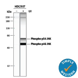
- Detection of Human Phospho-JNK (T183/Y185) by Simple WesternTM. Simple Western lane view shows lysates of HEK293T human embryonic kidney cell line untreated (-) or treated (+) with 100 J/m2 ultraviolet light (UV) for 30 minutes, loaded at 0.2 mg/mL. Specific bands were detected for Phospho-JNK (T183/Y185) at approximately 56 and 46 kDa (as indicated) using 5 µg/mL of Rabbit Anti-Human/Mouse/Rat Phospho-JNK (T183/Y185) Antigen Affinity-purified Polyclonal Antibody (Catalog # AF1205). This experiment was conducted under reducing conditions and using the 12-230 kDa separation system. *Non-specific interaction with the 230 kDa Simple Western standard may be seen with this antibody.

Supportive validation
- Submitted by
- Novus Biologicals (provider)
- Main image
- Experimental details
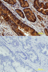
- JNK in Human Prostate. JNK phosphorylated at T183/Y185 was detected in immersion fixed paraffin-embedded sections of human prostate array using Rabbit Anti-Human/Mouse/Rat Phospho-JNK (T183/Y185) Antigen Affinity-purified Polyclonal Antibody (Catalog # AF1205) at 15 µg/mL overnight at 4 °C. Tissue was stained using the Anti-Rabbit HRP-DAB Cell & Tissue Staining Kit (brown; Catalog # CTS005) and counterstained with hematoxylin (blue). Lower panel shows a lack of labeling if primary antibodies are omitted and tissue is stained only with secondary antibody followed by incubation with detection reagents. View our protocol for Chromogenic IHC Staining of Paraffin-embedded Tissue Sections.

- Submitted by
- Novus Biologicals (provider)
- Main image
- Experimental details
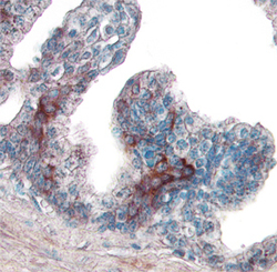
- JNK in Human Breast Cancer Tissue. JNK phosphorylated at T183/Y185 was detected in immersion fixed paraffin-embedded sections of human breast cancer tissue using Rabbit Anti-Human/Mouse/Rat Phospho-JNK (T183/Y185) Antigen Affinity-purified Polyclonal Antibody (Catalog # AF1205) at 15 µg/mL overnight at 4 °C. Tissue was stained using the Anti-Rabbit HRP-DAB Cell & Tissue Staining Kit (brown; Catalog # CTS005) and counterstained with hematoxylin (blue). View our protocol for Chromogenic IHC Staining of immersion fixed paraffin-embedded Tissue Sections.
