 Explore
 Validate
 Learn
Learn Western blot
 Immunocytochemistry
Antibody data
- Antibody Data
- Antigen structure
- References [3]
- Comments [0]
- Validations
- Western blot [2]
Submit
Validation data
Reference
Comment
Report error
- Product number
- MAB17761 - Provider product page

- Provider
- Novus Biologicals
- Product name
- Mouse Monoclonal JNK1 Antibody
- Antibody type
- Monoclonal
- Description
- Protein A or G purified from hybridoma culture supernatant. Detects human JNK1 in direct ELISAs and human, mouse, and rat JNK1 expressed predominantly as p46 JNK (isoforms 2 and/or 3, both 384 aa), with lesser amounts of p54 JNK (isoforms 1 and/or 4, both 427 aa), in Western blots. In Western blots, no cross-reactivity with recombinant human (rh) JNK2 or rhJNK3 is observed.
- Reactivity
- Human, Mouse, Rat
- Host
- Mouse
- Conjugate
- Unconjugated
- Isotype
- IgG
- Vial size
- 100 ug
- Concentration
- LYOPH
- Storage
- Use a manual defrost freezer and avoid repeated freeze-thaw cycles. 12 months from date of receipt, -20 to -70 degreesC as supplied. 1 month, 2 to 8 degreesC under sterile conditions after reconstitution. 6 months, -20 to -70 degreesC under sterile conditions after reconstitution.
Submitted references APP upregulation contributes to retinal ganglion cell degeneration via JNK3.
Phosphorylation Sites Identified in the NEIL1 DNA Glycosylase Are Potential Targets for the JNK1 Kinase.
Distinct role of c-Jun N-terminal kinase isoforms in human neutrophil apoptosis regulated by tumor necrosis factor-alpha and granulocyte-macrophage colony-stimulating factor.
Liu C, Zhang CW, Zhou Y, Wong WQ, Lee LC, Ong WY, Yoon SO, Hong W, Fu XY, Soong TW, Koo EH, Stanton LW, Lim KL, Xiao ZC, Dawe GS
Cell death and differentiation 2018 Mar;25(4):663-678
Cell death and differentiation 2018 Mar;25(4):663-678
Phosphorylation Sites Identified in the NEIL1 DNA Glycosylase Are Potential Targets for the JNK1 Kinase.
Prakash A, Cao VB, Doublié S
PloS one 2016;11(8):e0157860
PloS one 2016;11(8):e0157860
Distinct role of c-Jun N-terminal kinase isoforms in human neutrophil apoptosis regulated by tumor necrosis factor-alpha and granulocyte-macrophage colony-stimulating factor.
Kato T, Noma H, Kitagawa M, Takahashi T, Oshitani N, Kitagawa S
Journal of interferon & cytokine research : the official journal of the International Society for Interferon and Cytokine Research 2008 Apr;28(4):235-43
Journal of interferon & cytokine research : the official journal of the International Society for Interferon and Cytokine Research 2008 Apr;28(4):235-43
No comments: Submit comment
Supportive validation
- Submitted by
- Novus Biologicals (provider)
- Main image

- Experimental details
- Detection of Human, Mouse, and Rat JNK1 by Western Blot. Western blot shows lysates of CHP-100 human neuroblastoma cell line, C6 rat glioma cell line, and C2C12 mouse myoblast cell line. PVDF membrane was probed with 1 µg/mL Mouse Anti-Human/Mouse/Rat JNK1 Monoclonal Antibody (Catalog # MAB17761) followed by HRP-conjugated Anti-Mouse IgG Secondary Antibody (Catalog # HAF007). A specific band for JNK1 was detected at approximately 46 kDa and 54 kDa (as indicated). This experiment was conducted under reducing conditions and using Immunoblot Buffer Group 4.

- Submitted by
- Novus Biologicals (provider)
- Main image
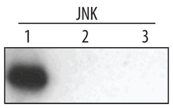
- Experimental details
- Detection of Human JNK1 by Western Blot. Western blot shows recombinant human JNK1, JNK2, and JNK3 (1 ng/lane). PVDF membrane was probed with 1 µg/mL Mouse Anti-Human/Mouse/Rat JNK1 Monoclonal Antibody (Catalog # MAB17761) followed by HRP-conjugated Anti-Mouse IgG Secondary Antibody (Catalog # HAF007). A specific band for JNK1 was detected at approximately 46 kDa (as indicated). This experiment was conducted under reducing conditions and using Immunoblot Buffer Group 4.
