Explore
 Validate
 Learn
Learn Western blot
 Immunohistochemistry
Antibody data
- Antibody Data
- Antigen structure
- References [16]
- Comments [0]
- Validations
- Immunohistochemistry [2]
- Flow cytometry [1]
- Other assay [5]
Submit
Validation data
Reference
Comment
Report error
- Product number
- 44-690G - Provider product page

- Provider
- Invitrogen Antibodies
- Product name
- JNK1 Polyclonal Antibody
- Antibody type
- Polyclonal
- Antigen
- Synthetic peptide
- Reactivity
- Human, Mouse, Rat
- Host
- Rabbit
- Isotype
- IgG
- Vial size
- 100 μL
- Storage
- -20°C
Submitted references Mechanical Study of Jian-Gan-Xiao-Zhi Decoction on Nonalcoholic Fatty Liver Disease Based on Integrated Network Pharmacology and Untargeted Metabolomics.
Heterogeneity in old fibroblasts is linked to variability in reprogramming and wound healing.
Expression of FADD and cFLIP(L) balances mitochondrial integrity and redox signaling to substantiate apoptotic cell death.
FADD regulates NF-κB activation and promotes ubiquitination of cFLIPL to induce apoptosis.
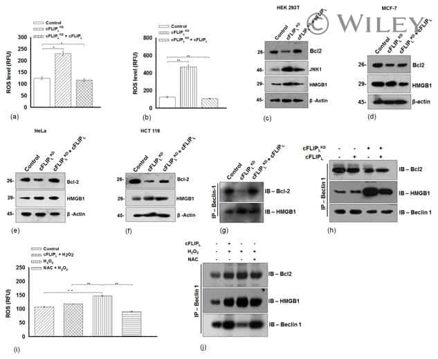
Expression of cFLIPL Determines the Basal Interaction of Bcl-2 With Beclin-1 and Regulates p53 Dependent Ubiquitination of Beclin-1 During Autophagic Stress.
Functional comparison of herpes simplex virus 1 (HSV-1) and HSV-2 ICP27 homologs reveals a role for ICP27 in virion release.
Imatinib restores VASP activity and its interaction with Zyxin in BCR-ABL leukemic cells.
Increased ERK and JNK activation and decreased ERK/JNK ratio are associated with long-term organ damage in patients with systemic lupus erythematosus.
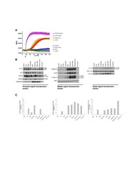
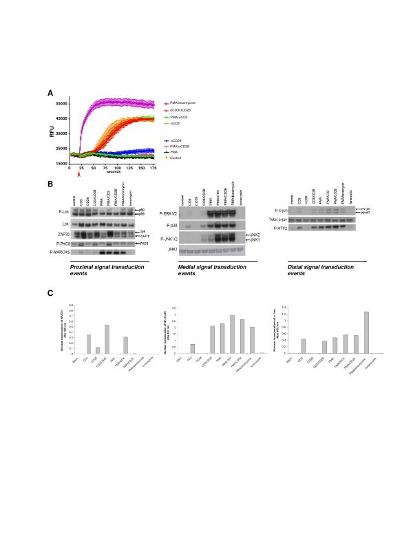
Molecular pathway profiling of T lymphocyte signal transduction pathways; Th1 and Th2 genomic fingerprints are defined by TCR and CD28-mediated signaling.
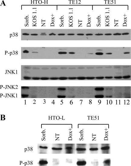
Herpes simplex virus type 1 ICP27 induces p38 mitogen-activated protein kinase signaling and apoptosis in HeLa cells.
Varicella-zoster virus infection of human fibroblast cells activates the c-Jun N-terminal kinase pathway.
The mitogen-activated protein kinases (MAPK) p38 and JNK are markers of tumor progression in breast carcinoma.
Homozygous deletion of MKK4 in ovarian serous carcinoma.
Homozygous deletion of MKK4 in ovarian serous carcinoma.
Matrix metalloproteinases (MMP), EMMPRIN (extracellular matrix metalloproteinase inducer) and mitogen-activated protein kinases (MAPK): co-expression in metastatic serous ovarian carcinoma.
Matrix metalloproteinases (MMP), EMMPRIN (extracellular matrix metalloproteinase inducer) and mitogen-activated protein kinases (MAPK): co-expression in metastatic serous ovarian carcinoma.
Cao YJ, Li HZ, Zhao J, Sun YM, Jin XW, Lv SQ, Luo JY, Fang XX, Wen WB, Liao JB
Evidence-based complementary and alternative medicine : eCAM 2022;2022:2264394
Evidence-based complementary and alternative medicine : eCAM 2022;2022:2264394
Heterogeneity in old fibroblasts is linked to variability in reprogramming and wound healing.
Mahmoudi S, Mancini E, Xu L, Moore A, Jahanbani F, Hebestreit K, Srinivasan R, Li X, Devarajan K, Prélot L, Ang CE, Shibuya Y, Benayoun BA, Chang ALS, Wernig M, Wysocka J, Longaker MT, Snyder MP, Brunet A
Nature 2019 Oct;574(7779):553-558
Nature 2019 Oct;574(7779):553-558
Expression of FADD and cFLIP(L) balances mitochondrial integrity and redox signaling to substantiate apoptotic cell death.
Ranjan K, Pathak C
Molecular and cellular biochemistry 2016 Nov;422(1-2):135-150
Molecular and cellular biochemistry 2016 Nov;422(1-2):135-150
FADD regulates NF-κB activation and promotes ubiquitination of cFLIPL to induce apoptosis.
Ranjan K, Pathak C
Scientific reports 2016 Mar 14;6:22787
Scientific reports 2016 Mar 14;6:22787
Expression of cFLIPL Determines the Basal Interaction of Bcl-2 With Beclin-1 and Regulates p53 Dependent Ubiquitination of Beclin-1 During Autophagic Stress.
Ranjan K, Pathak C
Journal of cellular biochemistry 2016 Aug;117(8):1757-68
Journal of cellular biochemistry 2016 Aug;117(8):1757-68
Functional comparison of herpes simplex virus 1 (HSV-1) and HSV-2 ICP27 homologs reveals a role for ICP27 in virion release.
Park D, Lalli J, Sedlackova-Slavikova L, Rice SA
Journal of virology 2015 Mar;89(5):2892-905
Journal of virology 2015 Mar;89(5):2892-905
Imatinib restores VASP activity and its interaction with Zyxin in BCR-ABL leukemic cells.
Bernusso VA, Machado-Neto JA, Pericole FV, Vieira KP, Duarte AS, Traina F, Hansen MD, Olalla Saad ST, Barcellos KS
Biochimica et biophysica acta 2015 Feb;1853(2):388-95
Biochimica et biophysica acta 2015 Feb;1853(2):388-95
Increased ERK and JNK activation and decreased ERK/JNK ratio are associated with long-term organ damage in patients with systemic lupus erythematosus.
Bloch O, Amit-Vazina M, Yona E, Molad Y, Rapoport MJ
Rheumatology (Oxford, England) 2014 Jun;53(6):1034-42
Rheumatology (Oxford, England) 2014 Jun;53(6):1034-42
Molecular pathway profiling of T lymphocyte signal transduction pathways; Th1 and Th2 genomic fingerprints are defined by TCR and CD28-mediated signaling.
Smeets RL, Fleuren WW, He X, Vink PM, Wijnands F, Gorecka M, Klop H, Bauerschmidt S, Garritsen A, Koenen HJ, Joosten I, Boots AM, Alkema W
BMC immunology 2012 Mar 14;13:12
BMC immunology 2012 Mar 14;13:12
Herpes simplex virus type 1 ICP27 induces p38 mitogen-activated protein kinase signaling and apoptosis in HeLa cells.
Gillis PA, Okagaki LH, Rice SA
Journal of virology 2009 Feb;83(4):1767-77
Journal of virology 2009 Feb;83(4):1767-77
Varicella-zoster virus infection of human fibroblast cells activates the c-Jun N-terminal kinase pathway.
Zapata HJ, Nakatsugawa M, Moffat JF
Journal of virology 2007 Jan;81(2):977-90
Journal of virology 2007 Jan;81(2):977-90
The mitogen-activated protein kinases (MAPK) p38 and JNK are markers of tumor progression in breast carcinoma.
Davidson B, Konstantinovsky S, Kleinberg L, Nguyen MT, Bassarova A, Kvalheim G, Nesland JM, Reich R
Gynecologic oncology 2006 Sep;102(3):453-61
Gynecologic oncology 2006 Sep;102(3):453-61
Homozygous deletion of MKK4 in ovarian serous carcinoma.
Nakayama K, Nakayama N, Davidson B, Katabuchi H, Kurman RJ, Velculescu VE, Shih IeM, Wang TL
Cancer biology & therapy 2006 Jun;5(6):630-4
Cancer biology & therapy 2006 Jun;5(6):630-4
Homozygous deletion of MKK4 in ovarian serous carcinoma.
Nakayama K, Nakayama N, Davidson B, Katabuchi H, Kurman RJ, Velculescu VE, Shih IeM, Wang TL
Cancer biology & therapy 2006 Jun;5(6):630-4
Cancer biology & therapy 2006 Jun;5(6):630-4
Matrix metalloproteinases (MMP), EMMPRIN (extracellular matrix metalloproteinase inducer) and mitogen-activated protein kinases (MAPK): co-expression in metastatic serous ovarian carcinoma.
Davidson B, Givant-Horwitz V, Lazarovici P, Risberg B, Nesland JM, Trope CG, Schaefer E, Reich R
Clinical & experimental metastasis 2003;20(7):621-31
Clinical & experimental metastasis 2003;20(7):621-31
No comments: Submit comment
Supportive validation
- Submitted by
- Invitrogen Antibodies (provider)
- Main image

- Experimental details
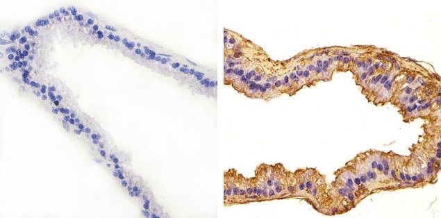
- Immunohistochemistry analysis of JNK1 showing staining in the cytoplasm of paraffin-embedded mouse prostate tissue (right) compared to a negative control without primary antibody (left). To expose target proteins, antigen retrieval was performed using 10mM sodium citrate (pH 6.0), microwaved for 8-15 min. Following antigen retrieval, tissues were blocked in 3% H2O2-methanol for 15 min at room temperature, washed with ddH2O and PBS, and then probed with a JNK1 Rabbit Polyclonal Antibody (Product # 44-690G) diluted in 3% BSA-PBS at a dilution of 1:100 overnight at 4°C in a humidified chamber. Tissues were washed extensively in PBST and detection was performed using an HRP-conjugated secondary antibody followed by colorimetric detection using a DAB kit. Tissues were counterstained with hematoxylin and dehydrated with ethanol and xylene to prep for mounting.
- Submitted by
- Invitrogen Antibodies (provider)
- Main image
- Experimental details
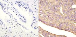
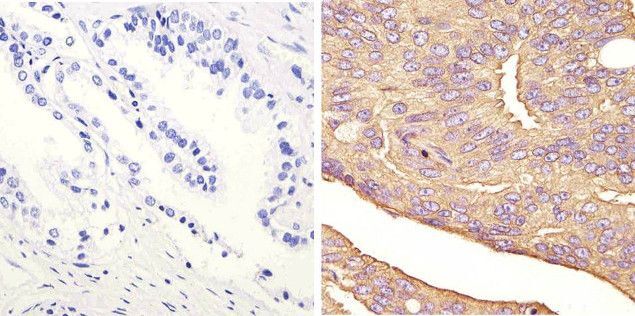
- Immunohistochemistry analysis of JNK1 showing staining in the cytoplasm of paraffin-embedded human prostate carcinoma (right) compared to a negative control without primary antibody (left). To expose target proteins, antigen retrieval was performed using 10mM sodium citrate (pH 6.0), microwaved for 8-15 min. Following antigen retrieval, tissues were blocked in 3% H2O2-methanol for 15 min at room temperature, washed with ddH2O and PBS, and then probed with a JNK1 Rabbit Polyclonal Antibody (Product # 44-690G) diluted in 3% BSA-PBS at a dilution of 1:100 overnight at 4°C in a humidified chamber. Tissues were washed extensively in PBST and detection was performed using an HRP-conjugated secondary antibody followed by colorimetric detection using a DAB kit. Tissues were counterstained with hematoxylin and dehydrated with ethanol and xylene to prep for mounting.
Supportive validation
- Submitted by
- Invitrogen Antibodies (provider)
- Main image
- Experimental details
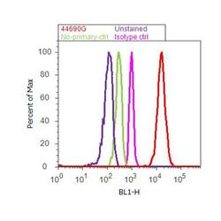
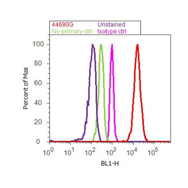
- Flow cytometry analysis of JNK1 was done on A-431. Cells were fixed with 70% ethanol for 10 minutes, permeabilized with 0.25% Triton™ X-100 for 20 minutes, and blocked with 5% BSA for 30 minutes at room temperature. Cells were labeled with JNK1 Rabbit Polyclonal Antibody (44690G, red histogram) or with rabbit isotype control (pink histogram) at 3-5 ug/million cells in 2.5% BSA. After incubation at room temperature for 2 hours, the cells were labeled with Alexa Fluor® 488 Goat Anti-Rabbit Secondary Antibody (A11008) at a dilution of 1:400 for 30 minutes at room temperature. The representative 10,000 cells were acquired and analyzed for each sample using an Attune® Acoustic Focusing Cytometer. The purple histogram represents unstained control cells and the green histogram represents no-primary-antibody control.
Supportive validation
- Submitted by
- Invitrogen Antibodies (provider)
- Main image
- Experimental details
- NULL

- Submitted by
- Invitrogen Antibodies (provider)
- Main image
- Experimental details
- NULL
- Submitted by
- Invitrogen Antibodies (provider)
- Main image

- Experimental details
- NULL
- Submitted by
- Invitrogen Antibodies (provider)
- Main image
- Experimental details
- Figure 1 Signal transduction in Jurkat T cells . Jurkat T cells were stimulated with different combinations of stimuli in order to elucidate the different signal transduction pathways. A ; Jurkat T cells were stimulated as indicated and intracellular Ca 2+ release was monitored over/in time. B ; Intracellular signal transduction routes were charted via phosphoanalysis using western blot. Jurkat T cells were stimulated for 15 min using different stimulations. Proximal (Lck, ZAP70, PKCtheta and the PKC substrate MARCKs), medial (MAPK phosphorylation) and distal (c-Jun and ATF2) signaling was monitored based on the phosphorylation status of the described proteins. C ; Nuclear translocation of the transcription factors NFAT, NFkB and c-Jun was evaluated 15 minutes after stimulation.
- Submitted by
- Invitrogen Antibodies (provider)
- Main image

- Experimental details
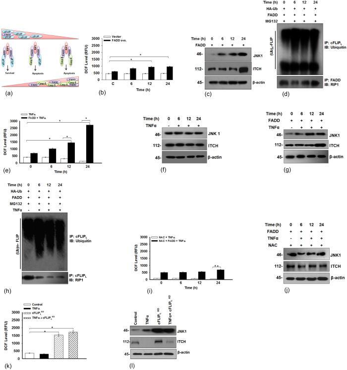
- Figure 6 FADD triggers JNK1 mediate ubiquitination of cFLIP L . ( a ) Illustration represents FADD stabilizes RIP1 and procaspase-8 associated pro-apoptotic complex II. ( b ) HEK 293T cells were transfected with pcDNA3.1-FADD for mentioned time points, control represents 48 h of FADD (shown as 0 h time point) expressed cells, to examine the cellular ROS, ( c ) Expression of JNK1 and ITCH and ( d ) The ubiquitination of cFLIP L in MG132 (10 muM for 3 h) pre-treated cells subjected to co-immunoprecipitation analysis. From the same cell lysate the co-imunoprecipitation of FADD with RIP1 was carried out. ( e ) HEK 293T cells were treated as mentioned in the figure legend 2c to examine the level of cellular ROS and ( f ) Expression of JNK1 and ITCH ( g ) HEK 293T cells were treated as mentioned in figure legend 2c and expression of JNK1 and ITCH ( h ) The 48 h pcDNA3.1-FADD transfected HEK 293T cells pre-treated with MG132 (10 muM for 3 h) followed by treatment of TNF-alpha (10 ng/ml) for mentioned time points and subjected to Co-IP assay for monitoring the ubiquitination of cFLIP L and expression of RIP1. ( i ) The 48 h pcDNA3.1-FADD transfected HEK 293T cells pre-treated with N-acetyl cysteine (NAC) (25 muM for 3 h) followed by treatment of TNF-alpha (10 ng/ml) for mentioned time points and level of cellular ROS and ( j ) Expression of JNK1 and ITCH ( k ) HEK 293T cells were treated as described above in the figure legend 2i to determine the level of cellular ROS and ( l ) Expre