 Explore
 Validate
 Learn
Learn Western blot
Antibody data
- Antibody Data
- Antigen structure
- References [4]
- Comments [0]
- Validations
- Western blot [1]
- Immunocytochemistry [1]
Submit
Validation data
Reference
Comment
Report error
- Product number
- GTX24821 - Provider product page

- Provider
- GeneTex
- Proper citation
- GeneTex Cat#GTX24821, RRID:AB_369523
- Product name
- JNK1 + JNK2 (phospho Thr183/Tyr185) antibody
- Antibody type
- Polyclonal
- Reactivity
- Human
- Host
- Rabbit
- Storage
- Aliquot and store at -20°C or -80¢XC. Avoid repeated freeze / thaw cycles.
Submitted references EGFR transactivation is involved in TNF-α-induced expression of thymic stromal lymphopoietin in human keratinocyte cell line.
The effects of artocarpin on wound healing: in vitro and in vivo studies.
Tanshinone IIA inhibits BT-20 human breast cancer cell proliferation through increasing caspase 12, GADD153 and phospho-p38 protein expression.
Reversine suppresses oral squamous cell carcinoma via cell cycle arrest and concomitantly apoptosis and autophagy.
Segawa R, Shigeeda K, Hatayama T, Dong J, Mizuno N, Moriya T, Hiratsuka M, Hirasawa N
Journal of dermatological science 2018 Mar;89(3):290-298
Journal of dermatological science 2018 Mar;89(3):290-298
The effects of artocarpin on wound healing: in vitro and in vivo studies.
Yeh CJ, Chen CC, Leu YL, Lin MW, Chiu MM, Wang SH
Scientific reports 2017 Nov 15;7(1):15599
Scientific reports 2017 Nov 15;7(1):15599
Tanshinone IIA inhibits BT-20 human breast cancer cell proliferation through increasing caspase 12, GADD153 and phospho-p38 protein expression.
Yan MY, Chien SY, Kuo SJ, Chen DR, Su CC
International journal of molecular medicine 2012 May;29(5):855-63
International journal of molecular medicine 2012 May;29(5):855-63
Reversine suppresses oral squamous cell carcinoma via cell cycle arrest and concomitantly apoptosis and autophagy.
Lee YR, Wu WC, Ji WT, Chen JY, Cheng YP, Chiang MK, Chen HR
Journal of biomedical science 2012 Jan 27;19:9
Journal of biomedical science 2012 Jan 27;19:9
No comments: Submit comment
Supportive validation
- Submitted by
- GeneTex (provider)
- Main image

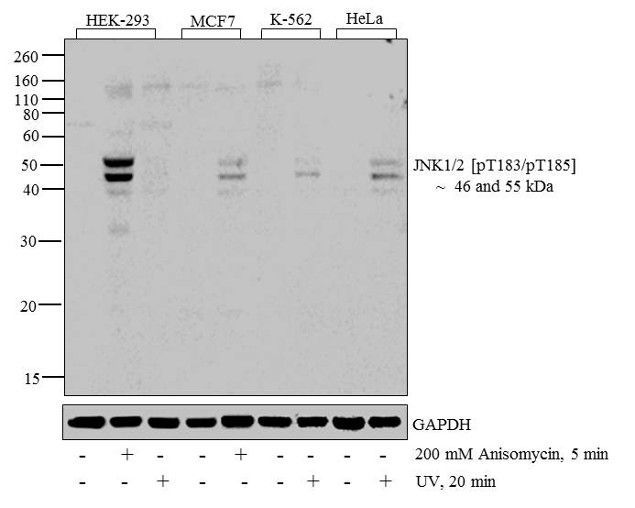
- Experimental details
- Western blot analysis of JNK1 + JNK2 [pT183 + pT185] was performed by loading 20 ?g of HEK-293 (lane1), HEK-293 treated for 5 minutes with 200 mM of Anisomycin (lane2), HEK-293 treated for 20 minutes with UV (lane3), MCF7 (lane4), MCF7 treated for 5 minutes with 200 mM of Anisomycin (lane5), K562 (lane6), K562 treated for 20 minutes with UV (lane7), HeLa (lane8) and HeLa treated for 20 minutes with UV (lane9) cell lysate using Novex??NuPAGE? ?4-12 % Bis-Tris gel, XCell SureLock? Electrophoresis System, Novex? Sharp Pre-Stained Protein Standard?, and iBlot? Dry Blotting System. Proteins were transferred to a nitrocellulose membrane and blocked with 5% skim milk for 1 hour at room temperature. JNK1 + JNK2 [pT183 + pT185] was detected at ~ 46 and 55 kDa using JNK1 + JNK2 [pT183 + pT185] Rabbit Polyclonal Antibody at 1:1000 dilution in 5% skim milk at 4¢XC overnight on a rocking platform. Goat Anti-Rabbit IgG - HRP Secondary Antibody at 1:5000 dilution was used and chemiluminescent detection was performed using Novex? ECL Chemiluminescent Substrate Reagent.
Supportive validation
- Submitted by
- GeneTex (provider)
- Main image

- Experimental details
- Immunofluorescence analysis of JNK1/2 [pT183/pT185] was done on 70% confluent log phase A549 cells treated with Anisomycin (25 £gg/mL for 30 min). The cells were fixed with 4% paraformaldehyde for 15 minutes, permeabilized with 0.25% Triton? X-100 for 10 minutes, and blocked with 5% BSA for 1 hour at room temperature. The cells were labeled with JNK1/2 [pT183/pT185] Rabbit polyclonal Antibody at 2 £gg/mL in 1% BSA and incubated for 3 hours at room temperature and then labeled with Alexa Flour 488 Goat Anti-Rabbit IgG Secondary Antibody at a dilution of 1:400 for 30 minutes at room temperature (Panel a: green). Nuclei (Panel b: blue) were stained with SlowFadeR Gold Antifade Mountant with DAPI. F-actin (Panel c: red) was stained with Alexa Fluor 594 Phalloidin. Panel d is a merged image showing translocation of JNK1/2 [pT183/pT185] to the nucleus upon Anisomycin treatment. Panel e is untreated cells showing cytoplasmic localization. Panel f shows no primary antibody control. The images were captured at 20X magnification.
