Explore
 Validate
 Learn
Learn Western blot
 ELISA
ELISA Immunocytochemistry
Antibody data
- Antibody Data
- Antigen structure
- References [5]
- Comments [0]
- Validations
- Immunocytochemistry [2]
- Immunohistochemistry [1]
- Flow cytometry [2]
- Other assay [1]
Submit
Validation data
Reference
Comment
Report error
- Product number
- MA5-15873 - Provider product page

- Provider
- Invitrogen Antibodies
- Product name
- Ku80 Monoclonal Antibody (5C5)
- Antibody type
- Monoclonal
- Antigen
- Purifed from natural sources
- Description
- MA5-15873 targets XRCC5 in indirect ELISA, FACS, IF, IHC, and WB applications and shows reactivity with Human and mouse samples. The MA5-15873 immunogen is purified recombinant fragment of human XRCC5 expressed in E. Coli. . MA5-15873 detects XRCC5 which has a predicted molecular weight of approximately 86kDa.
- Reactivity
- Human, Mouse
- Host
- Mouse
- Isotype
- IgG
- Antibody clone number
- 5C5
- Vial size
- 100 μL
- Concentration
- Conc. Not Determined
- Storage
- Store at 4°C short term. For long term storage, store at -20°C, avoiding freeze/thaw cycles.
Submitted references CAPRI enables comparison of evolutionarily conserved RNA interacting regions.
Lysine-specific demethylase 2A enhances binding of various nuclear factors to CpG-rich genomic DNAs by action of its CXXC-PHD domain.
Tyrosine kinase inhibitor-induced defects in DNA repair sensitize FLT3(ITD)-positive leukemia cells to PARP1 inhibitors.
Gene expression and mutation-guided synthetic lethality eradicates proliferating and quiescent leukemia cells.
IGH/MYC Translocation Associates with BRCA2 Deficiency and Synthetic Lethality to PARP1 Inhibitors.
Panhale A, Richter FM, Ramírez F, Shvedunova M, Manke T, Mittler G, Akhtar A
Nature communications 2019 Jun 18;10(1):2682
Nature communications 2019 Jun 18;10(1):2682
Lysine-specific demethylase 2A enhances binding of various nuclear factors to CpG-rich genomic DNAs by action of its CXXC-PHD domain.
Iuchi S, Paulo JA
Scientific reports 2019 Apr 2;9(1):5496
Scientific reports 2019 Apr 2;9(1):5496
Tyrosine kinase inhibitor-induced defects in DNA repair sensitize FLT3(ITD)-positive leukemia cells to PARP1 inhibitors.
Maifrede S, Nieborowska-Skorska M, Sullivan-Reed K, Dasgupta Y, Podszywalow-Bartnicka P, Le BV, Solecka M, Lian Z, Belyaeva EA, Nersesyan A, Machnicki MM, Toma M, Chatain N, Rydzanicz M, Zhao H, Jelinek J, Piwocka K, Sliwinski T, Stoklosa T, Ploski R, Fischer T, Sykes SM, Koschmieder S, Bullinger L, Valent P, Wasik MA, Huang J, Skorski T
Blood 2018 Jul 5;132(1):67-77
Blood 2018 Jul 5;132(1):67-77
Gene expression and mutation-guided synthetic lethality eradicates proliferating and quiescent leukemia cells.
Nieborowska-Skorska M, Sullivan K, Dasgupta Y, Podszywalow-Bartnicka P, Hoser G, Maifrede S, Martinez E, Di Marcantonio D, Bolton-Gillespie E, Cramer-Morales K, Lee J, Li M, Slupianek A, Gritsyuk D, Cerny-Reiterer S, Seferynska I, Stoklosa T, Bullinger L, Zhao H, Gorbunova V, Piwocka K, Valent P, Civin CI, Muschen M, Dick JE, Wang JC, Bhatia S, Bhatia R, Eppert K, Minden MD, Sykes SM, Skorski T
The Journal of clinical investigation 2017 Jun 1;127(6):2392-2406
The Journal of clinical investigation 2017 Jun 1;127(6):2392-2406
IGH/MYC Translocation Associates with BRCA2 Deficiency and Synthetic Lethality to PARP1 Inhibitors.
Maifrede S, Martin K, Podszywalow-Bartnicka P, Sullivan-Reed K, Langer SK, Nejati R, Dasgupta Y, Hulse M, Gritsyuk D, Nieborowska-Skorska M, Lupey-Green LN, Zhao H, Piwocka K, Wasik MA, Tempera I, Skorski T
Molecular cancer research : MCR 2017 Aug;15(8):967-972
Molecular cancer research : MCR 2017 Aug;15(8):967-972
No comments: Submit comment
Supportive validation
- Submitted by
- Invitrogen Antibodies (provider)
- Main image

- Experimental details
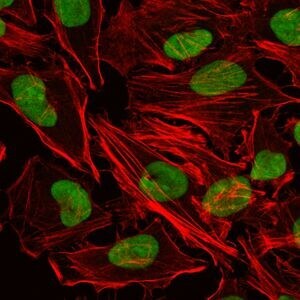
- Immunofluorescence analysis of HeLa cells using XRCC5 monoclonal antibody (Product # MA5-15873) (Green). Red: actin filaments have been labeled with phalloidin.
- Submitted by
- Invitrogen Antibodies (provider)
- Main image
- Experimental details
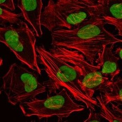
- Immunofluorescence analysis of HeLa cells using XRCC5 monoclonal antibody (Product # MA5-15873) (Green). Red: actin filaments have been labeled with phalloidin.

Supportive validation
- Submitted by
- Invitrogen Antibodies (provider)
- Main image
- Experimental details
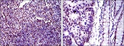
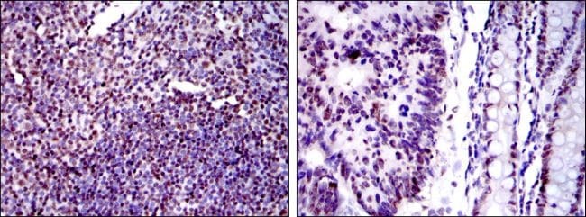
- Immunohistochemical analysis of paraffin-embedded human tonsil tissues (left) and human colon cancer tissues (right) using XRCC5 monoclonal antibody (Product # MA5-15873) followed with DAB staining.
Supportive validation
- Submitted by
- Invitrogen Antibodies (provider)
- Main image

- Experimental details
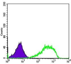
- Flow cytometric analysis of HeLa cells using XRCC5 monoclonal antibody (Product # MA5-15873) (green) and negative control (purple).

- Submitted by
- Invitrogen Antibodies (provider)
- Main image

- Experimental details
- Flow cytometric analysis of HeLa cells using XRCC5 monoclonal antibody (Product # MA5-15873) (green) and negative control (purple).
Supportive validation
- Submitted by
- Invitrogen Antibodies (provider)
- Main image

- Experimental details
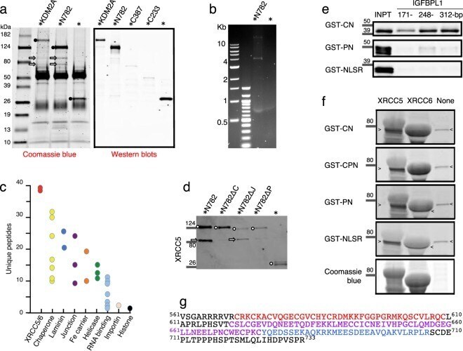
- Figure 2 Interaction of N782 with the XRCC5/6 complex. * Indicates EGFP tag. ( a ) Left panel shows proteins immunoprecipitated with a rabbit anti-GFP antibody (black circle) and co-immunoprecipitated proteins (arrow). It contains both rabbit IgG heavy chain (50 kD wide band) and the light chain (25 kD wide band) derived from the anti-GFP antibody. Right panel shows Western blots of immunoprecipitated EGFP-KDM2A, EGFP-N782, and EGFP-KDM2A C-termini. The proteins were detected by a combination of mouse anti-GFP (primary) and goat anti-mouse IgG. ( b ) Keratinocyte genomic DNA fragments co-immunoprecipitated with EGFP-N782 and EGFP. A marker (250 or 500 ng) or sample (5 mul) was applied to each well, respectively. ( c ) Mass spectrometry of proteins included in the 70-90 kDa protein bands [proteins from top to bottom, (XRCC5 and XRCC6 in red) and (TMPO and LMNA in blue)]. ( d ) Western blotting analysis of XRCC5 (arrow) co-immunoprecipitated with N782 or the mutants (circle): Delta = deletion, C = CXXC, J = JmjC, and P = PHD. ( e ) Binding of GST-CN, GST-PN, and GST-NLSR to 3-length IGFBPL1 baits. Full-length blots are presented in Supplementary Fig. S1 . ( f ) Far Western blotting analysis of the interaction of KMD2A domains with XRCC5 and 6. Probe and protein dye are on the left; cross-reaction of antibody to an E. coli protein, > and