Explore
 Validate
 Learn
Learn Western blot
Antibody data
- Antibody Data
- Antigen structure
- References [1]
- Comments [0]
- Validations
- Western blot [2]
- Immunocytochemistry [1]
- Immunohistochemistry [1]
- Other assay [1]
Submit
Validation data
Reference
Comment
Report error
- Product number
- PA5-27344 - Provider product page

- Provider
- Invitrogen Antibodies
- Product name
- MEF2A Polyclonal Antibody
- Antibody type
- Polyclonal
- Antigen
- Recombinant full-length protein
- Description
- Recommended positive controls: HeLa. Predicted reactivity: Mouse (100%), Rat (100%), Pig (99%), Bovine (100%). Store product as a concentrated solution. Centrifuge briefly prior to opening the vial.
- Reactivity
- Human, Mouse, Rat, Zebrafish
- Host
- Rabbit
- Isotype
- IgG
- Vial size
- 100 μL
- Concentration
- 0.77 mg/mL
- Storage
- Store at 4°C short term. For long term storage, store at -20°C, avoiding freeze/thaw cycles.
Submitted references Dysregulation of Muscle-Specific MicroRNAs as Common Pathogenic Feature Associated with Muscle Atrophy in ALS, SMA and SBMA: Evidence from Animal Models and Human Patients.
Malacarne C, Galbiati M, Giagnorio E, Cavalcante P, Salerno F, Andreetta F, Cagnoli C, Taiana M, Nizzardo M, Corti S, Pensato V, Venerando A, Gellera C, Fenu S, Pareyson D, Masson R, Maggi L, Dalla Bella E, Lauria G, Mantegazza R, Bernasconi P, Poletti A, Bonanno S, Marcuzzo S
International journal of molecular sciences 2021 May 26;22(11)
International journal of molecular sciences 2021 May 26;22(11)
No comments: Submit comment
Supportive validation
- Submitted by
- Invitrogen Antibodies (provider)
- Main image

- Experimental details
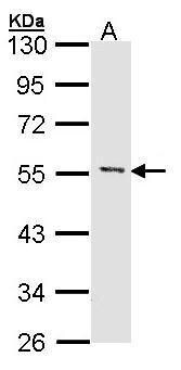
- Western Blot using MEF2A Polyclonal Antibody (Product # PA5-27344). Sample (30 µg of whole cell lysate). Lane A: Hela. 10% SDS PAGE. MEF2A Polyclonal Antibody (Product # PA5-27344) diluted at 1:1,000.
- Submitted by
- Invitrogen Antibodies (provider)
- Main image
- Experimental details
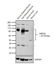
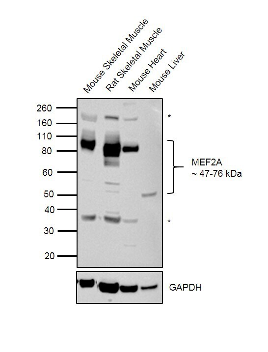
- Western blot was performed using Anti-MEF2A Rabbit Polyclonal Antibody (Product # PA5-27344) and 47-76 kDa bands corresponding to canonical as well as SUMOylated MEF2A isoforms were observed in tissues tested except Mouse Liver which is reported to be negative. Uncharacterized bands (*) were observed at around 38 kDa and 200 kDa. Whole cell extracts (30 µg lysate) of Mouse Skeletal Muscle (Lane 1), Rat Skeletal muscle (Lane 2), Mouse Heart (Lane 3) and Mouse Liver (Lane 4) were electrophoresed using Novex® NuPAGE® 4-12% Bis-Tris Protein Gel (Product # NP0322BOX). Resolved proteins were then transferred onto a nitrocellulose membrane (Product # IB23001) by iBlot® 2 Dry Blotting System (Product # IB21001). The blot was probed with the primary antibody (1:1000 dilution) and detected by Goat anti-Rabbit IgG (Heavy Chain) Superclonal™ Recombinant Secondary Antibody, HRP (Product # A27036, 1:4000 dilution) using the iBright FL 1000 (Product # A32752). Chemiluminescent detection was performed using Novex® ECL Chemiluminescent Substrate Reagent Kit (Product # WP20005)..
Supportive validation
- Submitted by
- Invitrogen Antibodies (provider)
- Main image
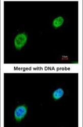
- Experimental details
- Immunofluorescent analysis of MEF2A in paraformaldehyde-fixed HeLa cells using a MEF2A polyclonal antibody (Product # PA5-27344) at a 1:200 dilution.

Supportive validation
- Submitted by
- Invitrogen Antibodies (provider)
- Main image

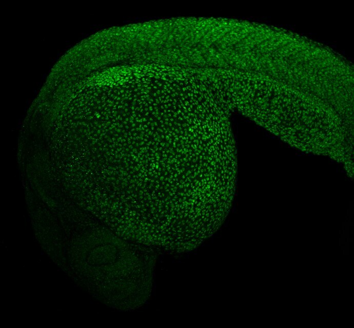
- Experimental details
- Immunohistochemistry (Free Floating) analysis of MEF2A was performed on paraformaldehyde-fixed 1 day-post-fertilization zebrafish embryo. Green: MEF2A was stained with MEF2A Polyclonal Antibody (Product # PA5-27344) diluted at 1:100.
Supportive validation
- Submitted by
- Invitrogen Antibodies (provider)
- Main image
- Experimental details
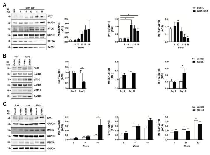
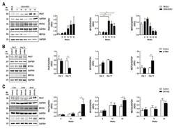
- Figure 3 Altered expression of PAX7, MYOG, and MEF2A proteins in G93A-SOD1, Delta7SMA, and AR113Q mouse muscle as disease progresses. Representative western blot analysis of PAX7, MYOG, and MEF2A proteins in gastrocnemius muscle tissue of ( A ) G93A-SOD1 (black bars), ( B ) Delta7SMA (black bars), and ( C ) AR113Q (black bars) mice and control mice (white bars) ( n = 3 mice per group) with relative densitometric analysis. Density values are reported as mean +- SEM, corrected for background and normalized to GAPDH control. * p < 0.05, Mann-Whitney test.