 Explore
 Validate
 Learn
Learn Western blot
Antibody data
- Antibody Data
- Antigen structure
- References [0]
- Comments [0]
- Validations
- Western blot [3]
- Immunohistochemistry [5]
- Flow cytometry [2]
Submit
Validation data
Reference
Comment
Report error
- Product number
- PA5-47027 - Provider product page

- Provider
- Invitrogen Antibodies
- Product name
- Neuropilin 1 Polyclonal Antibody
- Antibody type
- Polyclonal
- Antigen
- Recombinant full-length protein
- Description
- In direct ELISAs, approximately 25% cross-reactivity with recombinant human Neuropilin-1 is observed, and less than 1% cross-reactivity with recombinant human Neuropilin-2, recombinant mouse Neuropilin-2, and recombinant rat Neuropilin-2 is observed.
- Concentration
- 0.2 mg/mL
No comments: Submit comment
Supportive validation
- Submitted by
- Invitrogen Antibodies (provider)
- Main image
- Experimental details
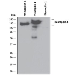
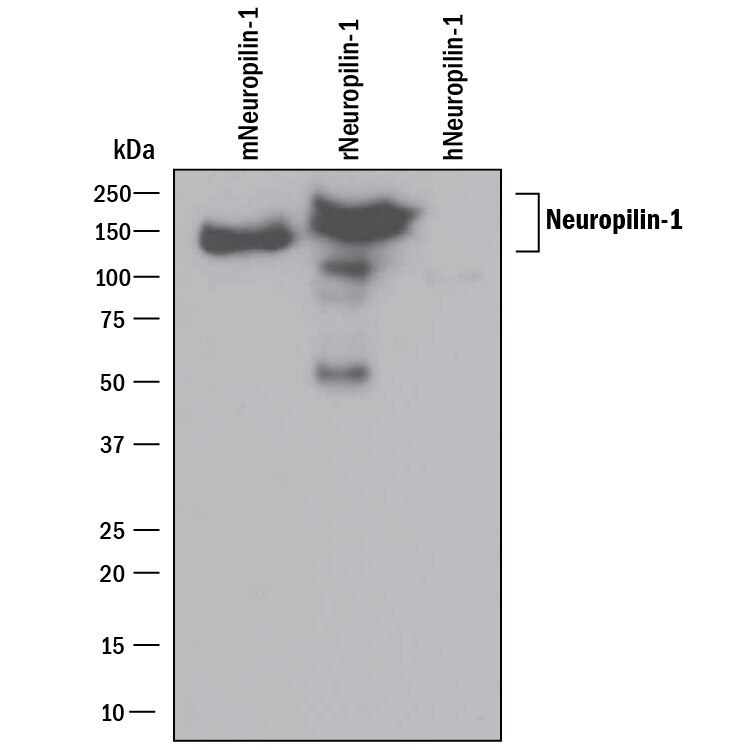
- Western blot analysis of Neuropilin 1 in 25 ng recombinant Mouse Neuropilin‚1, recombinant Rat Neuropilin‚1 Fc Chimera and recombinant Human Neuropilin‚1. Samples were incubated in Neuropilin 1 polyclonal antibody (Product # PA5-47027) using a dilution of 0.1 µg/mL followed by a HRP-conjugated Anti-Goat IgG secondary antibody. A specific band was detected for Neuropilin‚1 at approximately 150 kDa (as indicated). This experiment was conducted under reducing conditions.
- Submitted by
- Invitrogen Antibodies (provider)
- Main image

- Experimental details
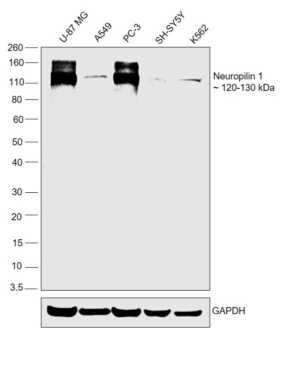
- Western blot was performed using Anti-Neuropilin 1 Polyclonal Antibody (Product # PA5-47027) and 120-130 kDa bands corresponding to Neuropilin 1 were observed to be higher in expression in U-87 MG, PC-3, and lower levels were observed in A549, SH-SY5Y and K562. Whole cell lysate (30ug lysate) of U-87 MG (Lane 1), A549 (Lane 2), PC-3 (Lane 3), SH-SY5Y (Lane 4) and K562 (Lane 5) were electrophoresed using Novex® NuPAGE® 4-12 % Bis-Tris gel (Product # NP0322BOX). Resolved proteins were then transferred onto a nitrocellulose membrane (Product # IB23001) by iBlot® 2 Dry Blotting System (Product # IB21001). The blot was probed with the primary antibody (0.1µg/ml) and detected by chemiluminescence with Rabbit anti-Goat IgG (H+L), Superclonal™ Recombinant Secondary Antibody, HRP (Product # A27014, 1:4000 dilution) using the iBright FL 1000 (Product # A32752). Chemiluminescent detection was performed using Novex® ECL Chemiluminescent Substrate Reagent Kit (Product # WP20005).
- Submitted by
- Invitrogen Antibodies (provider)
- Main image

- Experimental details
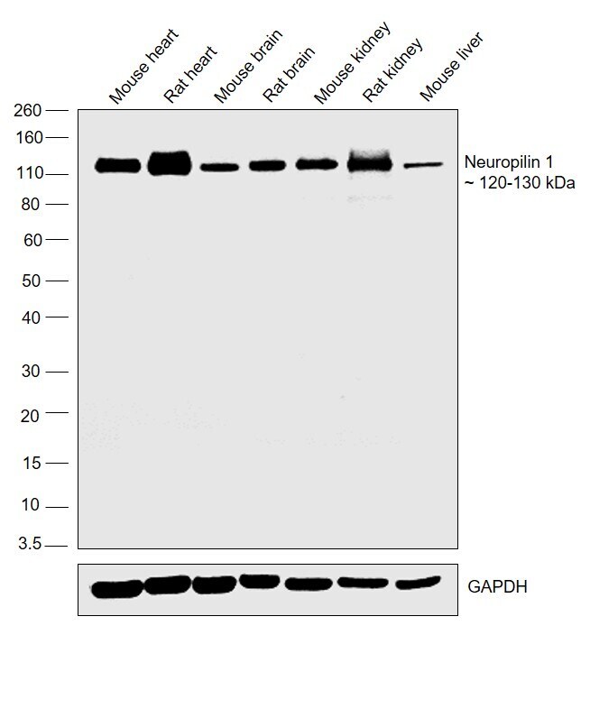
- Western blot was performed using Anti-Neuropilin 1 Polyclonal Antibody (Product # PA5-47027) and bands at 120-130 kDa band corresponding to Neuropilin 1 was observed to be higher in expression in Mouse Heart and Rat heart, and lower levels were observed in Mouse Brain, Rat Brain, Mouse Kidney, Rat Kidney and Mouse Liver. Whole cell lysate (30ug lysate) of Mouse heart (Lane 1), Rat heart (Lane 2), Mouse brain (Lane 3), Rat brain (Lane 4), Mouse kidney (Lane 5), Rat kidney (Lane 6) and Mouse liver (Lane 7) were electrophoresed using Novex® NuPAGE® 4-12 % Bis-Tris gel (Product # NP0322BOX). Resolved proteins were then transferred onto a nitrocellulose membrane (Product # IB23001) by iBlot® 2 Dry Blotting System (Product # IB21001). The blots were probed with the primary antibody (0.1µg/ml) and detected by chemiluminescence with Rabbit anti-Goat IgG (H+L), Superclonal™ Recombinant Secondary Antibody, HRP (Product # A27014, 1:4000 dilution) using the iBright FL 1000 (Product # A32752). Chemiluminescent detection was performed using Novex® ECL Chemiluminescent Substrate Reagent Kit (Product # WP20005).
Supportive validation
- Submitted by
- Invitrogen Antibodies (provider)
- Main image
- Experimental details
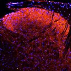
- Immunohistochemical analysis of Neuropilin 1 in perfusion fixed frozen sections of rat spinal cord. Samples were incubated with Neuropilin 1 polyclonal antibody (Product # PA5-47027) using a dilution of 15 µg/mL overnight at 4 °C followed by NorthernLights™ 557-conjugated Anti-Goat IgG Secondary Antibody (red) and counterstained with DAPI (blue). Specific staining was localized to the dorsal horn.

- Submitted by
- Invitrogen Antibodies (provider)
- Main image

- Experimental details
- Immunohistochemical analysis of Neuropilin 1 in perfusion fixed frozen sections of rat spinal cord. Samples were incubated with Neuropilin 1 polyclonal antibody (Product # PA5-47027) using a dilution of 15 µg/mL overnight at 4 °C followed by NorthernLights™ 557-conjugated Anti-Goat IgG Secondary Antibody (red) and counterstained with DAPI (blue). Specific staining was localized to the dorsal horn.

- Submitted by
- Invitrogen Antibodies (provider)
- Main image
- Experimental details
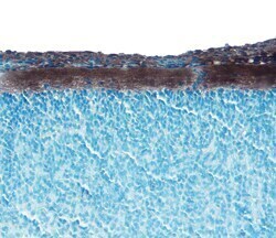
- Immunohistochemical analysis of Neuropilin 1 in immersion fixed frozen sections of embryonic rat spinal cord (15 d.p.c.). Samples were incubated in Neuropilin 1 polyclonal antibody (Product # PA5-47027) using a dilution of 5 µg/mL overnight at 4 °C. Tissue was stained with the Anti-Goat HRP-DAB Cell & Tissue Staining Kit (brown) and counterstained with hematoxylin (blue).

- Submitted by
- Invitrogen Antibodies (provider)
- Main image
- Experimental details
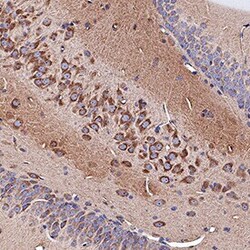
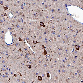
- Immunohistochemical analysis of Neuropilin 1 in immersion fixed paraffin-embedded sections of rat brain (hippocampus). Samples were incubated with Neuropilin 1 polyclonal antibody (Product # PA5-47027) using a dilution of 3 µg/mL for 1 hour at room temperature followed by Anti-Goat IgG VisUCyte™ HRP Polymer Antibody. Tissue was stained using DAB (brown) and counterstained with hematoxylin (blue). Specific staining was localized to cytoplasm in neuronal cell bodies and projections.
- Submitted by
- Invitrogen Antibodies (provider)
- Main image

- Experimental details
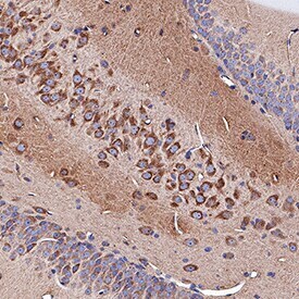
- Immunohistochemical analysis of Neuropilin 1 in immersion fixed paraffin-embedded sections of rat brain (thalamus). Samples were incubated with Neuropilin 1 polyclonal antibody (Product # PA5-47027) using a dilution of 3 µg/mL for 1 hour at room temperature followed by Anti-Goat IgG VisUCyte™ HRP Polymer Antibody. Tissue was stained using DAB (brown) and counterstained with hematoxylin (blue). Specific staining was localized to cytoplasm in neuronal cell bodies and projections.
Supportive validation
- Submitted by
- Invitrogen Antibodies (provider)
- Main image

- Experimental details
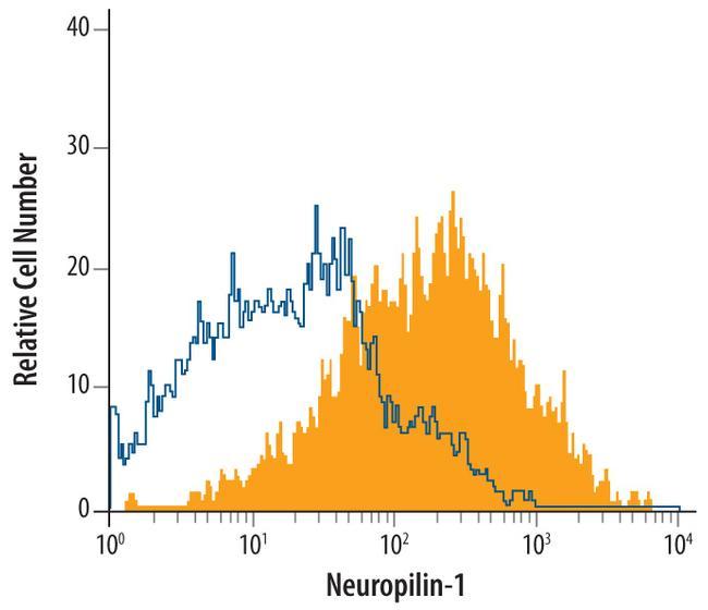
- Flow cytometric analysis of bEnd.3 mouse endothelioma cell line was stained with Goat Anti-Rat Neuropilin-1 Antigen Affinity-purified Polyclonal Antibody (Product # PA5-47027, filled histogram) or isotype control antibody (open histogram), followed by Allophycocyanin-conjugated Anti-Goat IgG Secondary Antibody.
- Submitted by
- Invitrogen Antibodies (provider)
- Main image

- Experimental details
- Flow cytometry of Neuropilin 1 in bEnd.3 mouse endothelioma cell line. Samples were incubated in Neuropilin 1 polyclonal antibody (Product # PA5-47027) or isotype control antibody followed by Allophycocyanin-conjugated Anti-Goat IgG Secondary Antibody.
