Explore
 Validate
 Learn
Learn Western blot
 Blocking/Neutralizing
Antibody data
- Antibody Data
- Antigen structure
- References [6]
- Comments [0]
- Validations
- Western blot [1]
- Immunocytochemistry [1]
- Immunohistochemistry [1]
- Flow cytometry [1]
Submit
Validation data
Reference
Comment
Report error
- Product number
- AF3870 - Provider product page

- Provider
- R&D Systems
- Product name
- Human Neuropilin-1 Antibody
- Antibody type
- Polyclonal
- Description
- Antigen Affinity-purified. Detects human Neuropilin-1 in direct ELISAs and Western blots. In direct ELISAs, less than 50% cross-reactivity with recombinant rat Neuropilin-1 and recombinant mouse Neuropilin-1 is observed, and less than 1% cross-reactivity with recombinant human Neuropilin-2 is observed.
- Reactivity
- Human
- Host
- Sheep
- Conjugate
- Unconjugated
- Antigen sequence
NP_001019799- Isotype
- IgG
- Vial size
- 100 ug
- Storage
- Use a manual defrost freezer and avoid repeated freeze-thaw cycles. 12 months from date of receipt, -20 to -70 °C as supplied. 1 month, 2 to 8 °C under sterile conditions after reconstitution. 6 months, -20 to -70 °C under sterile conditions after reconstitution.
Submitted references iRGD synergizes with PD-1 knockout immunotherapy by enhancing lymphocyte infiltration in gastric cancer.
Neuropilin-1 mediates neutrophil elastase uptake and cross-presentation in breast cancer cells.
Instruction of circulating endothelial progenitors in vitro towards specialized blood-brain barrier and arterial phenotypes.
Conformational remodeling of the fibronectin matrix selectively regulates VEGF signaling.
Neuropilin-1 mediates PDGF stimulation of vascular smooth muscle cell migration and signalling via p130Cas.
Alphav beta3 integrin limits the contribution of neuropilin-1 to vascular endothelial growth factor-induced angiogenesis.
Ding N, Zou Z, Sha H, Su S, Qian H, Meng F, Chen F, Du S, Zhou S, Chen H, Zhang L, Yang J, Wei J, Liu B
Nature communications 2019 Mar 22;10(1):1336
Nature communications 2019 Mar 22;10(1):1336
Neuropilin-1 mediates neutrophil elastase uptake and cross-presentation in breast cancer cells.
Kerros C, Tripathi SC, Zha D, Mehrens JM, Sergeeva A, Philips AV, Qiao N, Peters HL, Katayama H, Sukhumalchandra P, Ruisaard KE, Perakis AA, St John LS, Lu S, Mittendorf EA, Clise-Dwyer K, Herrmann AC, Alatrash G, Toniatti C, Hanash SM, Ma Q, Molldrem JJ
The Journal of biological chemistry 2017 Jun 16;292(24):10295-10305
The Journal of biological chemistry 2017 Jun 16;292(24):10295-10305
Instruction of circulating endothelial progenitors in vitro towards specialized blood-brain barrier and arterial phenotypes.
Boyer-Di Ponio J, El-Ayoubi F, Glacial F, Ganeshamoorthy K, Driancourt C, Godet M, Perrière N, Guillevic O, Couraud PO, Uzan G
PloS one 2014;9(1):e84179
PloS one 2014;9(1):e84179
Conformational remodeling of the fibronectin matrix selectively regulates VEGF signaling.
Ambesi A, McKeown-Longo PJ
Journal of cell science 2014 Sep 1;127(Pt 17):3805-16
Journal of cell science 2014 Sep 1;127(Pt 17):3805-16
Neuropilin-1 mediates PDGF stimulation of vascular smooth muscle cell migration and signalling via p130Cas.
Pellet-Many C, Frankel P, Evans IM, Herzog B, Jünemann-Ramírez M, Zachary IC
The Biochemical journal 2011 May 1;435(3):609-18
The Biochemical journal 2011 May 1;435(3):609-18
Alphav beta3 integrin limits the contribution of neuropilin-1 to vascular endothelial growth factor-induced angiogenesis.
Robinson SD, Reynolds LE, Kostourou V, Reynolds AR, da Silva RG, Tavora B, Baker M, Marshall JF, Hodivala-Dilke KM
The Journal of biological chemistry 2009 Dec 4;284(49):33966-81
The Journal of biological chemistry 2009 Dec 4;284(49):33966-81
No comments: Submit comment
Supportive validation
- Submitted by
- R&D Systems (provider)
- Main image
- Experimental details
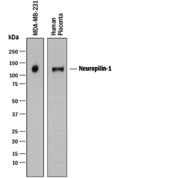
- Detection of Human Neuropilin-1 by Western Blot. Western blot shows lysates of MDA-MB-231 human breast cancer cell line and human placenta tissue. PVDF membrane was probed with 1 µg/mL of Sheep Anti-Human Neuropilin-1 Antigen Affinity-purified Polyclonal Antibody (Catalog # AF3870) followed by HRP-conjugated Anti-Sheep IgG Secondary Antibody (Catalog # HAF016). A specific band was detected for Neuropilin-1 at approximately 130 kDa (as indicated). This experiment was conducted under reducing conditions and using Immunoblot Buffer Group 1.
Supportive validation
- Submitted by
- R&D Systems (provider)
- Main image

- Experimental details
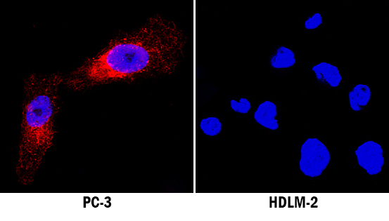
- Neuropilin-1 in PC-3 and HLDM-2 Human Cell Lines. Neuropilin-1 was detected in immersion fixed PC-3 human prostate cancer cell line (positive staining) and HLDM-2 human Hodgkin's lymphoma cell line (negative staining) using Sheep Anti-Human Neuropilin-1 Antigen Affinity-purified Polyclonal Antibody (Catalog # AF3870) at 5 µg/mL for 3 hours at room temperature. Cells were stained using the NorthernLights™ 557-conjugated Anti-Sheep IgG Secondary Antibody (red; Catalog # NL010) and counterstained with DAPI (blue). Specific staining was localized to cytoplasm. View our protocol for Fluorescent ICC Staining of Cells on Coverslips.
Supportive validation
- Submitted by
- R&D Systems (provider)
- Main image

- Experimental details
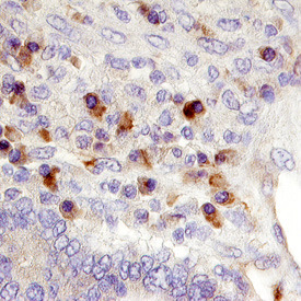
- Neuropilin-1 in Human Pancreatic Cancer Tissue. Neuropilin-1 was detected in immersion fixed paraffin-embedded sections of human pancreatic cancer tissue using Sheep Anti-Human Neuropilin-1 Antigen Affinity-purified Polyclonal Antibody (Catalog # AF3870) at 10 µg/mL overnight at 4 °C. Before incubation with the primary antibody tissue was subjected to heat-induced epitope retrieval using Antigen Retrieval Reagent-Basic (Catalog # CTS013). Tissue was stained using the Anti-Sheep HRP-DAB Cell & Tissue Staining Kit (brown; Catalog # CTS019) and counterstained with hematoxylin (blue). Specific labeling was localized to the cytoplasm and plasma membrane of cancer cells. View our protocol for Chromogenic IHC Staining of Paraffin-embedded Tissue Sections.
Supportive validation
- Submitted by
- R&D Systems (provider)
- Main image
- Experimental details
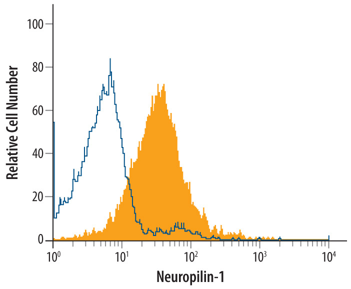
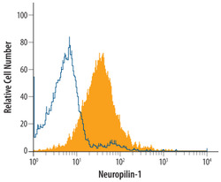
- Detection of Neuropilin-1 in HUVEC Human Cells by Flow Cytometry. HUVEC human umbilical vein endothelial cells were stained with Sheep Anti-Human Neuropilin-1 Antigen Affinity-purified Polyclonal Antibody (Catalog # AF3870, filled histogram) or control antibody (Catalog # 5-001-A, open histogram), followed by NorthernLights™ 637-conjugated Anti-Sheep IgG Secondary Antibody (Catalog # NL011).