Explore
 Validate
 Learn
Learn Western blot
Antibody data
- Antibody Data
- Antigen structure
- References [3]
- Comments [0]
- Validations
- Western blot [1]
Submit
Validation data
Reference
Comment
Report error
- Product number
- MAB3209 - Provider product page

- Provider
- Abnova Corporation
- Proper citation
- Abnova Corporation Cat#MAB3209, RRID:AB_10552878
- Product name
- NFATC1 monoclonal antibody, clone AT1C3
- Antibody type
- Monoclonal
- Description
- Mouse monoclonal antibody raised against partial recombinant NFATC1.
- Isotype
- IgG
- Antibody clone number
- AT1C3
- Storage
- Store at -20°C.Aliquot to avoid repeated freezing and thawing.
Submitted references Epigenetic changes and suppression of the nuclear factor of activated T cell 1 (NFATC1) promoter in human lymphomas with defects in immunoreceptor signaling.
Autoamplification of NFATc1 expression determines its essential role in bone homeostasis.
Transcription factors of the NFAT family: regulation and function.
Akimzhanov A, Krenacs L, Schlegel T, Klein-Hessling S, Bagdi E, Stelkovics E, Kondo E, Chuvpilo S, Wilke P, Avots A, Gattenlöhner S, Müller-Hermelink HK, Palmetshofer A, Serfling E
The American journal of pathology 2008 Jan;172(1):215-24
The American journal of pathology 2008 Jan;172(1):215-24
Autoamplification of NFATc1 expression determines its essential role in bone homeostasis.
Asagiri M, Sato K, Usami T, Ochi S, Nishina H, Yoshida H, Morita I, Wagner EF, Mak TW, Serfling E, Takayanagi H
The Journal of experimental medicine 2005 Nov 7;202(9):1261-9
The Journal of experimental medicine 2005 Nov 7;202(9):1261-9
Transcription factors of the NFAT family: regulation and function.
Rao A, Luo C, Hogan PG
Annual review of immunology 1997;15:707-47
Annual review of immunology 1997;15:707-47
No comments: Submit comment
Supportive validation
- Submitted by
- Abnova Corporation (provider)
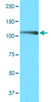
- Main image

- Experimental details
- Cell lysates of Ramos (20 ug) were resolved by SDS-PAGE, transferred to NC membrane and probed with NFATC1 monoclonal antibody, clone AT1C3 (Cat # MAB3209 ; 1 : 1000). Proteins were visualized using a goat anti-mouse secondary antibody conjugated to HRP and an ECL detection system.