 Explore
 Validate
 Learn
Learn Western blot
 Immunohistochemistry
Antibody data
- Antibody Data
- Antigen structure
- References [1]
- Comments [0]
- Validations
- Immunohistochemistry [3]
- Other assay [1]
Submit
Validation data
Reference
Comment
Report error
- Product number
- PA5-100732 - Provider product page

- Provider
- Invitrogen Antibodies
- Product name
- NIK Polyclonal Antibody
- Antibody type
- Polyclonal
- Antigen
- Synthetic peptide
- Description
- Antibody detects endogenous levels of total NIK.
- Reactivity
- Human, Mouse
- Host
- Rabbit
- Isotype
- IgG
- Vial size
- 100 μL
- Concentration
- 1 mg/mL
- Storage
- -20°C
Submitted references Therapeutic Benefit of Galectin-1: Beyond Membrane Repair, a Multifaceted Approach to LGMD2B.
Vallecillo-Zúniga ML, Poulson PD, Luddington JS, Arnold CJ, Rathgeber M, Kartchner BC, Hayes S, Gill H, Valdoz JC, Spallino JL, Garfield S, Dodson EL, Arthur CM, Stowell SR, Van Ry PM
Cells 2021 Nov 17;10(11)
Cells 2021 Nov 17;10(11)
No comments: Submit comment
Supportive validation
- Submitted by
- Invitrogen Antibodies (provider)
- Main image

- Experimental details
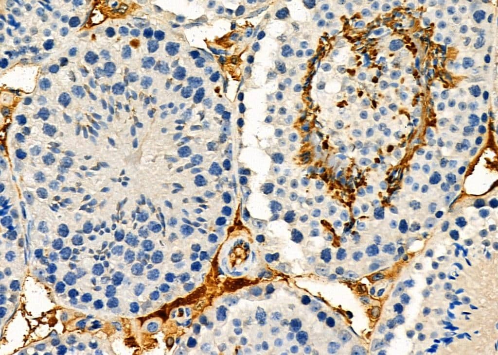
- Immunohistochemistry analysis of NIK in mouse testis tissue. The sample was formaldehyde fixed and a heat mediated antigen retrieval step in citrate buffer was performed. Samples were incubated with NIK polyclonal antibody (Product # PA5-100732) using a dilution of 1:100 (4°C overnight) followed by HRP conjugated anti-Rabbit secondary antibody.
- Submitted by
- Invitrogen Antibodies (provider)
- Main image

- Experimental details
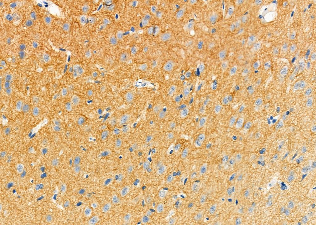
- Immunohistochemistry analysis of NIK in rat brain tissue. The sample was formaldehyde fixed and a heat mediated antigen retrieval step in citrate buffer was performed. Samples were incubated with NIK polyclonal antibody (Product # PA5-100732) using a dilution of 1:100 (4°C overnight) followed by HRP conjugated anti-Rabbit secondary antibody.
- Submitted by
- Invitrogen Antibodies (provider)
- Main image

- Experimental details
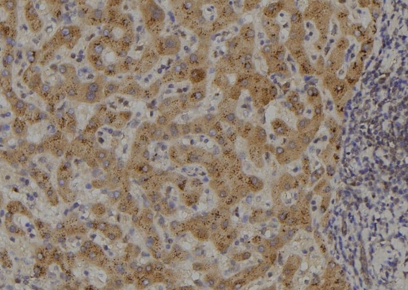
- Immunohistochemistry analysis of paraffin-embedded NIK in human liver tissue. Antigen retrieval was performed using citrate buffer. Samples were blocked with blocking buffer (1.5 hr, 22°C), incubated with NIK polyclonal antibody (Product # PA5-100732) using a dilution of 1:100 (1.5 hr, 22°C), followed by HRP conjugated goat anti-rabbit.
Supportive validation
- Submitted by
- Invitrogen Antibodies (provider)
- Main image
- Experimental details
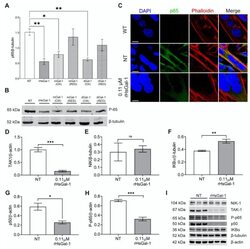
- Figure 2 In vitro treatment with rHsGal-1 modulates inflammatory response through the NF-kappaB pathway. ( A ) Quantification of expression levels of p65 (normalized to beta-tubulin) in 48 h A/J -/- NT or 0.11 muM rHsGal-1 treated myotubes. ( B ) Western blot images showing the p65 expression in NT or 0.11 muM rHsGal-1 48 h A/J -/- treated myotubes. ( C ) Representative images of WT and NT or 0.11 muM rHsGal-1 48 h A/J -/- treated myotubes cultured and immunostained with p65 (green), Phalloidin (red), and DAPI (blue). ( D - H ) Western blot quantification of 48 h NT or 0.11 muM rHsGal-1treated myotubes expressing levels of TAK1 ( D ), NIK ( E ), IKBalpha ( F ), p50 ( G ), and P-p65 ( H ). ( I ) Western blot images of 48 h NT or 0.11 muM rHsGal-1treated myotubes expressing NF-kappaB inflammatory subunits quantified in D-H. n = 3 in each group. A. * p < 0.05 and ** p < 0.01 NT vs. all forms of Gal-1. D-H. * p < 0.05, ** p < 0.01, *** p < 0.001 NT vs. rHsGal-1. Data are represented as SEM.
