 Explore
 Validate
 Learn
Learn Western blot
 Immunocytochemistry
Antibody data
- Antibody Data
- Antigen structure
- References [1]
- Comments [0]
- Validations
- Immunocytochemistry [5]
- Immunohistochemistry [1]
- Flow cytometry [2]
- Other assay [1]
Submit
Validation data
Reference
Comment
Report error
- Product number
- PA1-9073 - Provider product page

- Provider
- Invitrogen Antibodies
- Product name
- p47phox Polyclonal Antibody
- Antibody type
- Polyclonal
- Antigen
- Synthetic peptide
- Description
- This antibody is predicted to react with bovine, mouse, porcine, rabbit and rat based on sequence homology. This antibody is tested in Peptide ELISA: antibody detection limit dilution 20,000.
- Reactivity
- Human, Mouse, Porcine
- Host
- Goat
- Isotype
- IgG
- Vial size
- 100 μg
- Concentration
- 0.5 mg/mL
- Storage
- -20°C, Avoid Freeze/Thaw Cycles
Submitted references TSPO ligand FGIN-1-27 controls priapism in sickle cell mice via endogenous testosterone production.
Musicki B, Karakus S, La Favor JD, Chen H, Silva FH, Sturny M, Zirkin BR, Burnett AL
Journal of cellular physiology 2021 Apr;236(4):3073-3082
Journal of cellular physiology 2021 Apr;236(4):3073-3082
No comments: Submit comment
Supportive validation
- Submitted by
- Invitrogen Antibodies (provider)
- Main image

- Experimental details
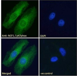
- Immunocytochemical analysis of p47phox in HeLa cells using a p47phox polyclonal antibody (Product #PA1-9073). HeLa cells were permeabilized with 0.15% Triton. Cells were incubated with 10 µg/mL of primary antibody for one hour followed by an Alexa Fluor 488 secondary antibody at a concentration of 2 µg/mL. Cytoplasmic staining can be seen as shown above. The nuclear stain is DAPI (blue). Negative control: Unimmunized goat IgG (10 µg/mL) followed by an Alexa Fluor 488 secondary antibody (2 µg/mL).

- Submitted by
- Invitrogen Antibodies (provider)
- Main image
- Experimental details
- Immunocytochemical analysis of p47phox in HeLa cells using a p47phox polyclonal antibody (Product #PA1-9073). HeLa cells were permeabilized with 0.15% Triton. Cells were incubated with 10 µg/mL of primary antibody for one hour followed by an Alexa Fluor 488 secondary antibody at a concentration of 2 µg/mL. Cytoplasmic staining can be seen as shown above. The nuclear stain is DAPI (blue). Negative control: Unimmunized goat IgG (10 µg/mL) followed by an Alexa Fluor 488 secondary antibody (2 µg/mL).

- Submitted by
- Invitrogen Antibodies (provider)
- Main image

- Experimental details
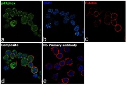
- Immunofluorescence analysis of p47phox was performed using 70% confluent log phase Raji cells. The cells were fixed with 4% paraformaldehyde for 10 minutes, permeabilized with 0.1% Triton™ X-100 for 15 minutes, and blocked with 2% BSA for 1 hour at room temperature. The cells were labeled with p47phox Goat Polyclonal Antibody (Product # PA1-9073) at 5 µg/mL in 0.1% BSA, incubated at 4 degree Celsius overnight and then labeled with Rabbit anti-Goat IgG (H+L) Cross-Adsorbed Secondary Antibody, Alexa Fluor 488 (Product # A-11078) at a dilution of 1:2000 for 45 minutes at room temperature (Panel a: green). Nuclei (Panel b: blue) were stained with ProLong™ Diamond Antifade Mountant with DAPI (Product # P36962). F-actin (Panel c: red) was stained with Rhodamine Phalloidin (Product # R415). Panel d represents the merged image showing cytoplasmic and membrane localization. Panel e represents control cells with no primary antibody to assess background. The images were captured at 60X magnification.

- Submitted by
- Invitrogen Antibodies (provider)
- Main image
- Experimental details
- Immunofluorescence analysis of p47phox was performed using 70% confluent log phase Raji cells. The cells were fixed with 4% paraformaldehyde for 10 minutes, permeabilized with 0.1% Triton™ X-100 for 15 minutes, and blocked with 2% BSA for 1 hour at room temperature. The cells were labeled with p47phox Goat Polyclonal Antibody (Product # PA1-9073) at 5 µg/mL in 0.1% BSA, incubated at 4 degree Celsius overnight and then labeled with Rabbit anti-Goat IgG (H+L) Cross-Adsorbed Secondary Antibody, Alexa Fluor 488 (Product # A-11078) at a dilution of 1:2000 for 45 minutes at room temperature (Panel a: green). Nuclei (Panel b: blue) were stained with ProLong™ Diamond Antifade Mountant with DAPI (Product # P36962). F-actin (Panel c: red) was stained with Rhodamine Phalloidin (Product # R415). Panel d represents the merged image showing cytoplasmic and membrane localization. Panel e represents control cells with no primary antibody to assess background. The images were captured at 60X magnification.

- Submitted by
- Invitrogen Antibodies (provider)
- Main image

- Experimental details
- Immunocytochemical analysis of p47phox in HeLa cells using a p47phox polyclonal antibody (Product #PA1-9073). HeLa cells were permeabilized with 0.15% Triton. Cells were incubated with 10 µg/mL of primary antibody for one hour followed by an Alexa Fluor 488 secondary antibody at a concentration of 2 µg/mL. Cytoplasmic staining can be seen as shown above. The nuclear stain is DAPI (blue). Negative control: Unimmunized goat IgG (10 µg/mL) followed by an Alexa Fluor 488 secondary antibody (2 µg/mL).

Supportive validation
- Submitted by
- Invitrogen Antibodies (provider)
- Main image

- Experimental details
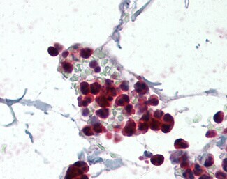
- Immunohistochemistry analysis of p47phox in human colon, neutrophils. Samples were incubated with p47phox polyclonal antibody (Product # PA1-9073) using a dilution of 5 µg/mL. Formalin-fixed, paraffin-embedded tissue after heat-induced antigen retrieval.
Supportive validation
- Submitted by
- Invitrogen Antibodies (provider)
- Main image

- Experimental details
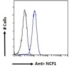
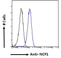
- Flow cytometric analysis of p47phox in HeLa cells using a polyclonal antibody (Product #PA1-9073). HeLa cells (blue line) were paraformaldehyde fixed and permeabilized with 0.5% Triton. The primary antibody was incubated for one hour (10 µg/mL) followed by an Alexa Fluor 488 secondary antibody (1 µg/mL). IgG control: Unimmunized goat IgG (black line) followed by an Alexa Fluor 488 secondary antibody.

- Submitted by
- Invitrogen Antibodies (provider)
- Main image
- Experimental details
- Flow cytometric analysis of p47phox in HeLa cells using a polyclonal antibody (Product #PA1-9073). HeLa cells (blue line) were paraformaldehyde fixed and permeabilized with 0.5% Triton. The primary antibody was incubated for one hour (10 µg/mL) followed by an Alexa Fluor 488 secondary antibody (1 µg/mL). IgG control: Unimmunized goat IgG (black line) followed by an Alexa Fluor 488 secondary antibody.
Supportive validation
- Submitted by
- Invitrogen Antibodies (provider)
- Main image
- Experimental details
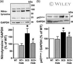
- 6 Figure Protein expressions of nitrotyrosine and p47phox subunit of NADPH oxidase were increased in the SCD compared to WT mouse penis and were decreased in the SCD mouse penis by FGIN-1-27 treatment. Representative western blots and quantification of nitrotyrosine (a) and p47phox (b) in penes isolated from WT and SCD mice treated with a TSPO ligand FGIN-1-27 or vehicle for nine days. Protein levels of nitrotyrosine and p47phox were analyzed by immunoblotting and normalized to GAPDH expression. Statistical analysis was performed by using one-way analysis of variance, followed by Newman-Keuls multiple comparison tests (between treatment groups) and by modified t test (between control and each treatment group). Data represent mean +- SEM ( n = 5-6). * p < .05 versus WT; # p < .05 versus SCD. FGIN-1-27, N , N -dihexyl-2-(4-fluorophenyl)indole-3-acetamide; GAPDH, glyceraldehyde-3-phosphate dehydrogenase; SCD, sickle cell disease; TSPO, translocator protein; WT, wild-type
