Explore
 Validate
 Learn
Learn Western blot
 Immunocytochemistry
Antibody data
- Antibody Data
- Antigen structure
- References [1]
- Comments [0]
- Validations
- Immunocytochemistry [4]
- Immunohistochemistry [1]
- Other assay [1]
Submit
Validation data
Reference
Comment
Report error
- Product number
- PA5-27821 - Provider product page

- Provider
- Invitrogen Antibodies
- Product name
- p47phox Polyclonal Antibody
- Antibody type
- Polyclonal
- Antigen
- Recombinant full-length protein
- Description
- Recommended positive controls: Raji. Predicted reactivity: Human (99%), Mouse (83%), Rat (80%), Pig (82%), Rabbit (86%), Bovine (87%). Store product as a concentrated solution. Centrifuge briefly prior to opening the vial.
- Reactivity
- Human
- Host
- Rabbit
- Isotype
- IgG
- Vial size
- 100 μL
- Concentration
- 1 mg/mL
- Storage
- Store at 4°C short term. For long term storage, store at -20°C, avoiding freeze/thaw cycles.
Submitted references The Presence of Cholesteryl Ester Transfer Protein (CETP) in Endothelial Cells Generates Vascular Oxidative Stress and Endothelial Dysfunction.
Wanschel ACBA, Guizoni DM, Lorza-Gil E, Salerno AG, Paiva AA, Dorighello GG, Davel AP, Balkan W, Hare JM, Oliveira HCF
Biomolecules 2021 Jan 7;11(1)
Biomolecules 2021 Jan 7;11(1)
No comments: Submit comment
Supportive validation
- Submitted by
- Invitrogen Antibodies (provider)
- Main image

- Experimental details
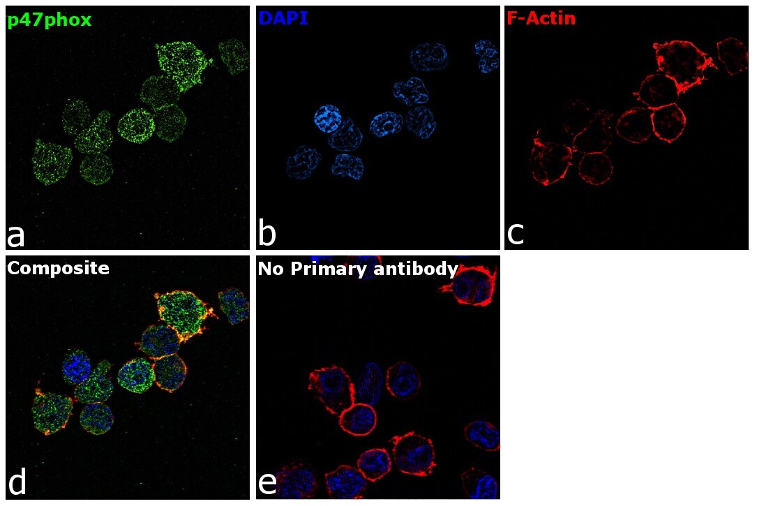
- p47phox Polyclonal Antibody detects NCF1 protein at cytoplasm by immunofluorescent analysis. Sample: Raji cells were fixed in 4% paraformaldehyde at RT for 15 min. Green: NCF1 protein stained by p47phox Polyclonal Antibody (Product # PA5-27821) diluted at 1:500. Blue: Hoechst 33342 staining.

- Submitted by
- Invitrogen Antibodies (provider)
- Main image

- Experimental details
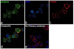
- Immunofluorescence analysis of p47phox was performed using 70% confluent log phase Raji cells. The cells were fixed with 4% paraformaldehyde for 10 minutes, permeabilized with 0.1% Triton™ X-100 for 15 minutes, and blocked with 2% BSA for 1 hour at room temperature. The cells were labeled with p47phox Rabbit Polyclonal Antibody (Product # PA5-27821) at 5 µg/mL in 0.1% BSA, incubated at 4 degree Celsius overnight and then with Goat anti-Rabbit IgG (H+L) Highly Cross-Adsorbed Secondary Antibody, Alexa Fluor Plus 488 (Product # A32731) at a dilution of 1:2000 for 45 minutes at room temperature (Panel a: green). Nuclei (Panel b: blue) were stained with ProLong™ Diamond Antifade Mountant with DAPI (Product # P36962). F-actin (Panel c: red) was stained with Rhodamine Phalloidin (Product # R415, 1:300). Panel d represents the merged image showing membrane localization. Panel e represents control cells with no primary antibody to assess background. The images were captured at 60X magnification.

- Submitted by
- Invitrogen Antibodies (provider)
- Main image
- Experimental details
- Immunofluorescence analysis of p47phox was performed using 70% confluent log phase Raji cells. The cells were fixed with 4% paraformaldehyde for 10 minutes, permeabilized with 0.1% Triton™ X-100 for 15 minutes, and blocked with 2% BSA for 1 hour at room temperature. The cells were labeled with p47phox Rabbit Polyclonal Antibody (Product # PA5-27821) at 5 µg/mL in 0.1% BSA, incubated at 4 degree Celsius overnight and then with Goat anti-Rabbit IgG (H+L) Highly Cross-Adsorbed Secondary Antibody, Alexa Fluor Plus 488 (Product # A32731) at a dilution of 1:2000 for 45 minutes at room temperature (Panel a: green). Nuclei (Panel b: blue) were stained with ProLong™ Diamond Antifade Mountant with DAPI (Product # P36962). F-actin (Panel c: red) was stained with Rhodamine Phalloidin (Product # R415, 1:300). Panel d represents the merged image showing membrane localization. Panel e represents control cells with no primary antibody to assess background. The images were captured at 60X magnification.
- Submitted by
- Invitrogen Antibodies (provider)
- Main image
- Experimental details
- p47phox Polyclonal Antibody detects NCF1 protein at cytoplasm by immunofluorescent analysis. Sample: Raji cells were fixed in 4% paraformaldehyde at RT for 15 min. Green: NCF1 protein stained by p47phox Polyclonal Antibody (Product # PA5-27821) diluted at 1:500. Blue: Hoechst 33342 staining.

Supportive validation
- Submitted by
- Invitrogen Antibodies (provider)
- Main image
- Experimental details
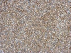
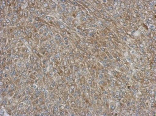
- Immunohistochemical analysis of paraffin-embedded Hela xenograft, using NCF1 (Product # PA5-27821) antibody at 1:500 dilution. Antigen Retrieval: EDTA based buffer, pH 8.0, 15 min.
Supportive validation
- Submitted by
- Invitrogen Antibodies (provider)
- Main image
- Experimental details
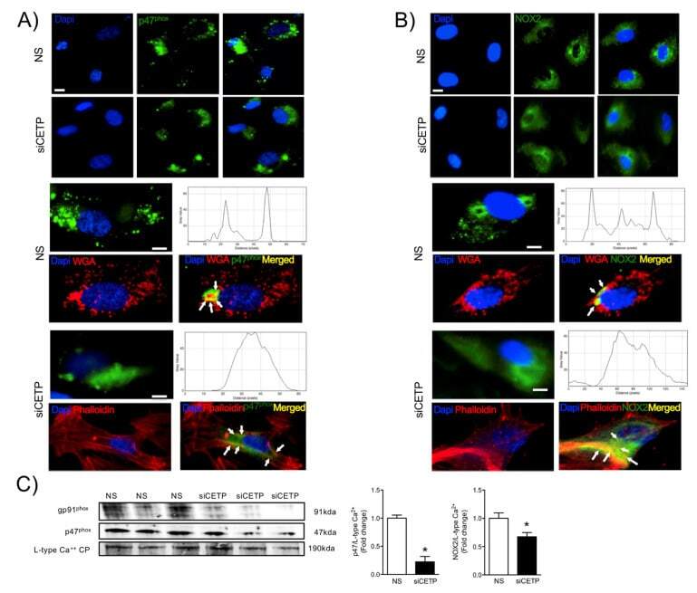
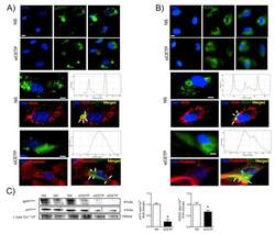
- Figure 4 CETP inhibition decreases NADPH oxidase activation. Representative microscopic images of HAEC stained with ( A ) p47 phox ( B ) NOX2 (gp91 phox ) and DAPI. p47 phox and NOX2 (green) are localized to the plasma membrane, stained with wheat germ agglutinin (WGA-red). After CETP inhibition, p47 phox and NOX2 co-localize with cytoplasmic aggregates of endogenous actin, detectable with phalloidin labeling. Upper panel 10X magnification and bottom 20X. Scale bar, 10 mum. ( C ) Western blotting of p47 phox and NOX2 (gp91 phox ) in the plasma membrane fraction of HAEC normalized by the L-type calcium channel protein ( n = 3, * p < 0.05). HAECs, human aortic endothelial cells. CETP, cholesteryl ester transfer protein. NS, non-silenced. siCETP, silenced CETP.