Explore
 Validate
 Learn
Learn Western blot
Antibody data
- Antibody Data
- Antigen structure
- References [4]
- Comments [0]
- Validations
- Western blot [1]
- Immunohistochemistry [1]
Submit
Validation data
Reference
Comment
Report error
- Product number
- sc-15340 - Provider product page

- Provider
- Santa Cruz Biotechnology
- Proper citation
- Santa Cruz Biotechnology Cat#sc-15340, RRID:AB_2237080
- Product name
- Anti-PIN1
- Antibody type
- Polyclonal
- Antigen
- Recombinant full-length protein
- Reactivity
- Human
- Host
- Rabbit
Submitted references Elevated PIN1 expression by C/EBPalpha-p30 blocks C/EBPalpha-induced granulocytic differentiation through c-Jun in AML.
Pin1 facilitates the phosphorylation-dependent ubiquitination of SF-1 to regulate gonadotropin beta-subunit gene transcription.
The prolyl isomerase Pin1 regulates the NF-kappaB signaling pathway and interleukin-8 expression in glioblastoma.
Peptidyl-prolyl isomerase Pin1 markedly enhances the oncogenic activity of the rel proteins in the nuclear factor-kappaB family.
Pulikkan JA, Dengler V, Peer Zada AA, Kawasaki A, Geletu M, Pasalic Z, Bohlander SK, Ryo A, Tenen DG, Behre G
Leukemia 2010 May;24(5):914-23
Leukemia 2010 May;24(5):914-23
Pin1 facilitates the phosphorylation-dependent ubiquitination of SF-1 to regulate gonadotropin beta-subunit gene transcription.
Luo Z, Wijeweera A, Oh Y, Liou YC, Melamed P
Molecular and cellular biology 2010 Feb;30(3):745-63
Molecular and cellular biology 2010 Feb;30(3):745-63
The prolyl isomerase Pin1 regulates the NF-kappaB signaling pathway and interleukin-8 expression in glioblastoma.
Atkinson GP, Nozell SE, Harrison DK, Stonecypher MS, Chen D, Benveniste EN
Oncogene 2009 Oct 22;28(42):3735-45
Oncogene 2009 Oct 22;28(42):3735-45
Peptidyl-prolyl isomerase Pin1 markedly enhances the oncogenic activity of the rel proteins in the nuclear factor-kappaB family.
Fan G, Fan Y, Gupta N, Matsuura I, Liu F, Zhou XZ, Lu KP, Gélinas C
Cancer research 2009 Jun 1;69(11):4589-97
Cancer research 2009 Jun 1;69(11):4589-97
No comments: Submit comment
Supportive validation
- Submitted by
- per
- Main image
- Experimental details
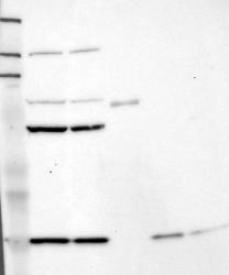
- Western blot analysis of antibody specificity using a routine panel composed of IgG/HSA-depleted human plasma and protein lysates from selected human tissues and cell lines.
- Validation comment
- Band of predicted size in kDa (+/-20%) with additional bands present.
- Primary Ab dilution
- 1:500
- Secondary Ab dilution
- 1:3000
- Lane 1
- Marker [kDa]: 219, 111, 83, 48, 32, 26, 17
- Lane 2
- RT-4
- Lane 3
- U-251MG sp
- Lane 4
- Human Plasma
- Lane 5
- Liver
- Lane 6
- Tonsil
- Theoretical target weight
- [kDa] 5

Supportive validation
- Submitted by
- per
- Main image

- Experimental details
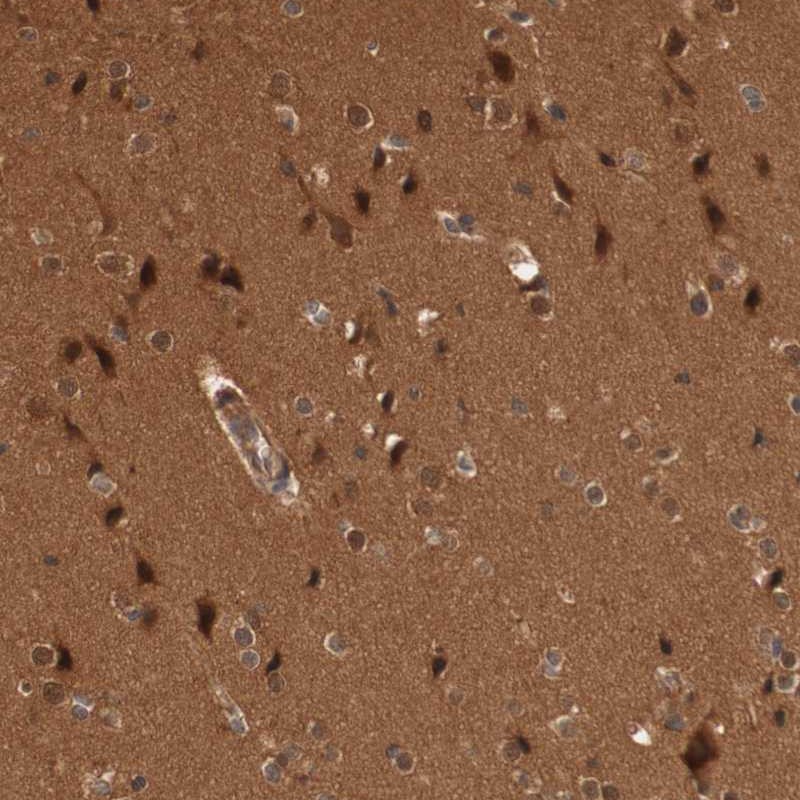
- Immunohistochemical staining of human cerebral cortex shows distinct cytoplasmic and nuclear positivity in neuronal cells.
- Validation comment
- Staining pattern partly consistent with experimental and/or bioinformatic data.